
|
|
The shape of the nodule - case 2025
|
|


Clinical data: A 21-yr-old woman was referred for evaluation of a newly diagnosed hypothyroidism. She was investigated because of fatigue.
Palpation: no abnormality.
Laboratory test: TSH 7.19 mIU/L, aTPO > 1300 U/mL.
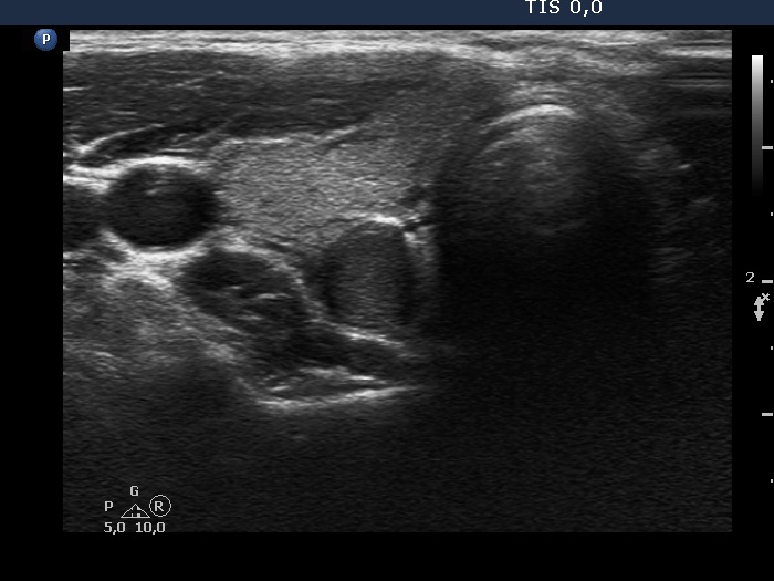
Ultrasonography. The thyroid was echonormal and had numerous discrete hypoechoic areas. The largest one located in the dorsal part seemed to be a pathological nodule at first sight. This lesion showed taller-than-wide shape and had blurred borders. Considering the presence of other, similarly hypoechoic areas in the thyroid, this lesion may be also the presentation of the underlying thyroiditis.
Cytology of the lesion in the right lobe resulted in Hashimoto's thyroiditis.
Comment. This is not an infrequent situation that we cannot decide whether a discrete lesion is a nodule in pathological sense or not. In this case, the blurred borders stand against a pathological nodule, while the relatively large size stands for this possibility. This dilemma is mainly theoretical and should not influence the decision on FNA.