
|
|
The shape of the nodule - case 2052
|
|

Clinical data: A 61-yr-old woman was referred for evaluation of a nodular goiter which was discovered on carotid Doppler examination.
Palpation: no abnormality.
Laboratory test: TSH 1.37 mIU/L.
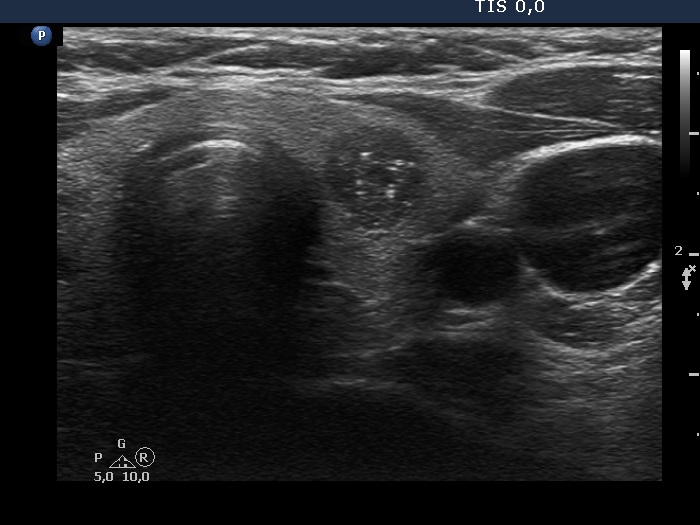
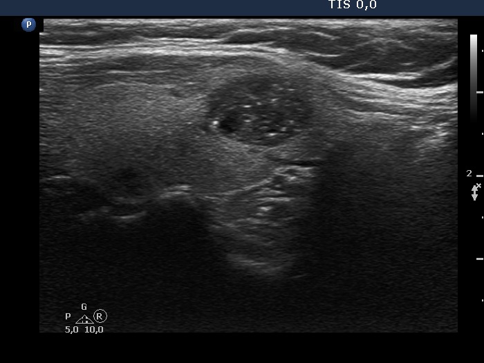
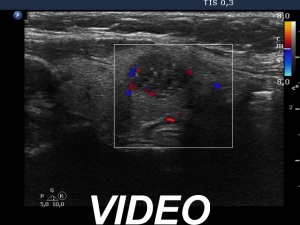
Ultrasonography. The thyroid was echonormal. There was hypoechoic nodule in the left lobe. The nodule had numerous echogenic granules and lines. Video record proved that these are clearly related to ventral cystic areas, therefore these are back wall cystic figures caused by posterior enhancement. The nodule showed taller-than-wide sign.
Aspiration cytology resulted in benign, cystic-colloid goiter.
Suggestion ultrasound in 3 to 5 years.
Comment. This case illustrates why is video clearly superior to still images in analyzing thyroid nodules. Viewing the still image on transverse section (first image in the list), the intranodular echogenic figures seem to be microcalcification. However, video clearly proved the presence of tiny cystic areas ventral to these echogenic figures.