
|
|
Graves' disease - case 121
|
|

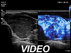
Right lobe, transverse scan


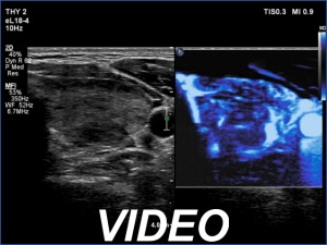
Right lobe, longitudinal scan


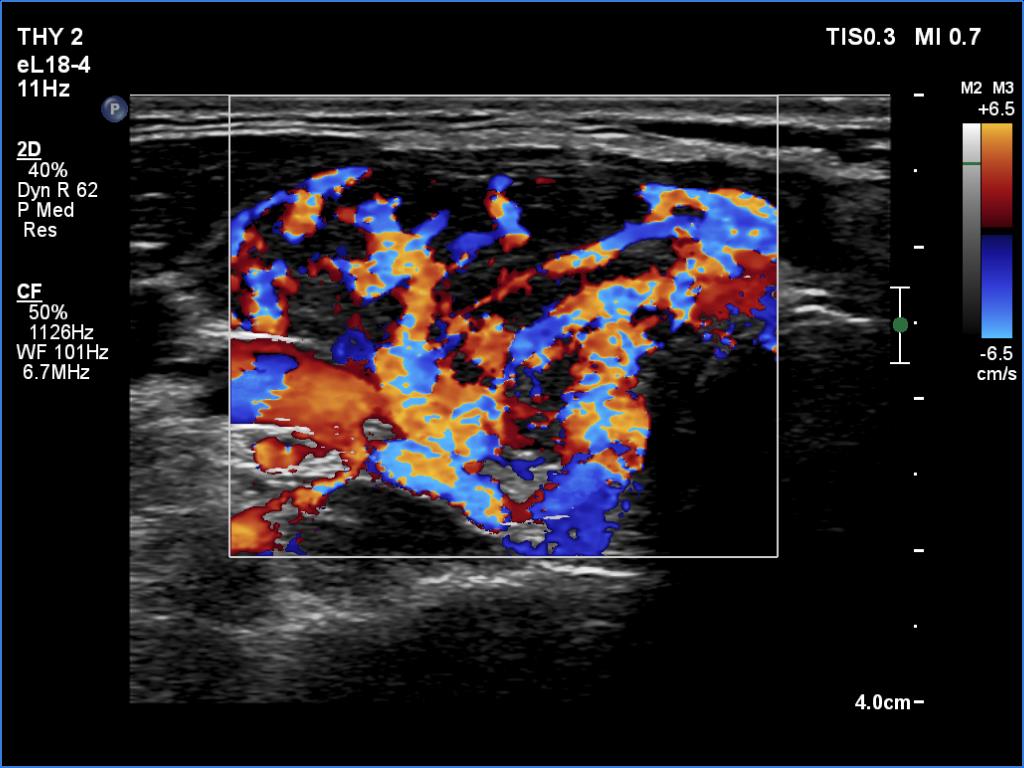
Right lobe, color Doppler mode


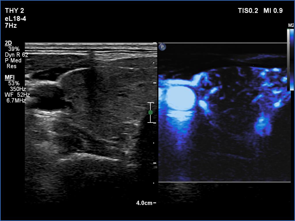
Right lobe, microflow imaging


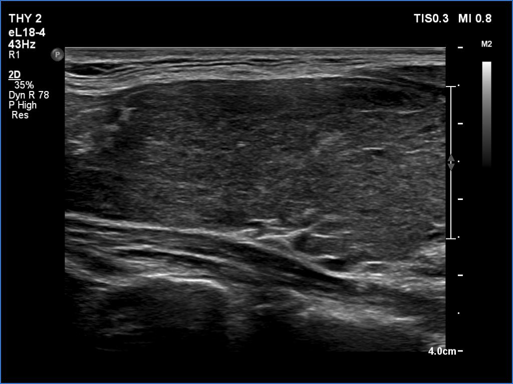
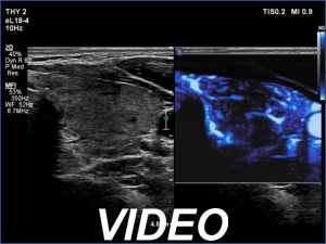
Left lobe, transverse scan


Left lobe, longitudinal scan


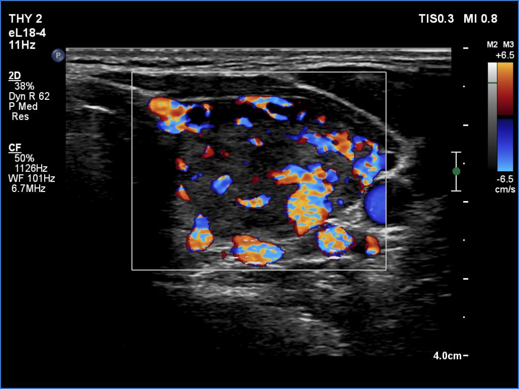
Left lobe, color Doppler mode


Left lobe, microflow imaging


Clinical data: A 35-year-old woman was referred for a newly diagnosed hyperthyroidism. The patient had typical complaints, including elevated heart rate and weight loss.
Summary of ultrasound and laboratory data:
Date of examination |
Volume of the thyroid lobes (mL) |
Echogenicity index (%) |
Therapy before |
Results of laboratory investigations |
Therapy after |
|||
|
Right lobe |
Left lobe |
TSH (mIU/L) |
FT4 (pM/L) |
FT3 (pM/L) |
||||
First visit |
27.2 |
18.4 |
70% |
Nothing |
< 0.001 |
61.2 |
35.7 |
30 mg methimazole |
5 weeks after the first visit |
19.1 |
13.5 |
50% |
30 mg methimazole |
< 0.001 |
7.85 |
6.19 |
15 mg methimazole |
4 months after the first visit |
10.4 |
14.4 |
20% |
15 mg methimazole |
0.06 |
13.7 |
4.61 |
10 mg methimazole |
9 months after the first visit |
13.9 |
12.0 |
10% |
10 mg methimazole |
1.98 |
8.11 |
- |
5 mg methimazole |
12 months after the first visit |
10.1 |
11.3 |
< 10% |
5 mg methimazole every other day |
4.97 |
10.1 |
- |
Nothing |
Comment.
This is the typical course of a non-relapsing Graves' hyperthyroidism. As the hormone results improve, both the size of the initially enlarged goiter and the degree of hypoechogenicity decrease.