
Graves' disease - case 2151
Three years after the first examination (ultrasonographic picture 5)
|
|

|
|
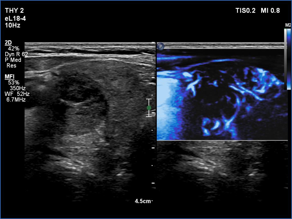
Right lobe, transverse scan, microflow imaging. The lesion has significantly fewer vessels than the extralesional parenchyma.