
|
|
Graves' disease - case 2151
|
|

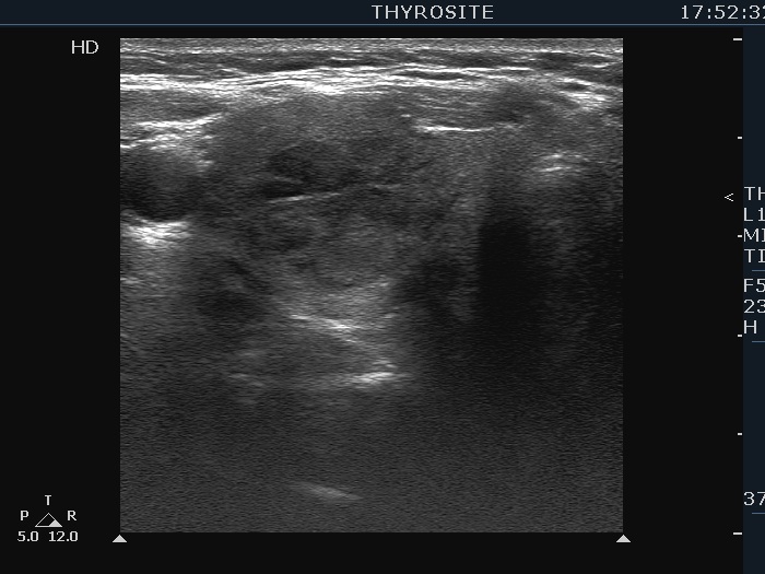

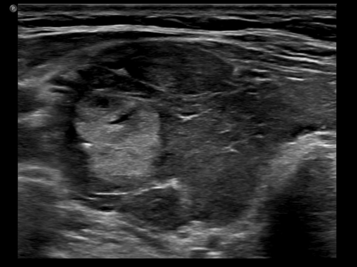


First examination (first row of images):
Clinical presentation: A 54-year-old woman was referred for an evaluation of complaints suggesting hyperthyroidism. She was treated with Graves' disease 16 years ago.
Palpation: Both lobes were enlarged and moderately firm.
Results of blood tests: hyperthyroidism (TSH undetectable, FT4 33.1 pM/L).
Ultrasonography. The thyroid was moderately hypoechoic. There was a more hypoechoic lesion in the central part of the lobe. The lesion was avascular.Aspiration cytology of the lesion resulted in benign hormonal atypia.
Thyrostatic therapy was started.
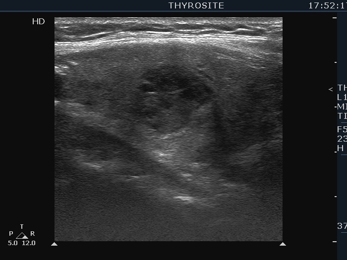
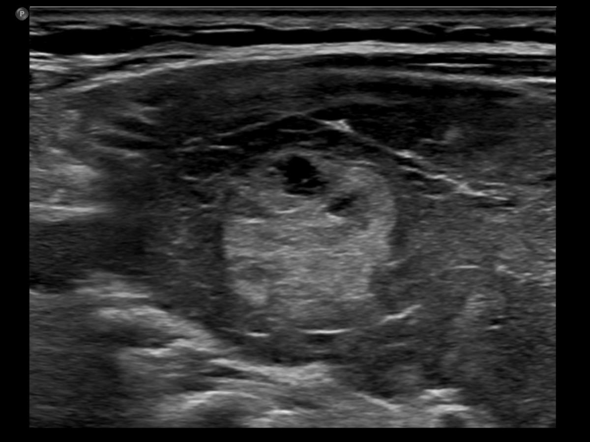
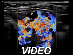
One year after the first examination (second row of images):
Clinical presentation. The patient had no complaints She came to follow-up visit.
Palpation: unchanged.
Results of blood tests: subclinical hyperthyroidism on daily 10 mg methimazole (TSH 0.001 mIU/L, FT4 13.8 pM/L, TSAb 8.2 U/L (normal value below 1.5)).
Ultrasonography. The thyroid became more hypoechoic while the lesion in the central part of the right lobe did hyperechoic. The vascularization of the thyroid was extremely increased.The patient was told that it is very likely that her hyperthyroidism will relapse therefore we suggested radioiodine therapy.
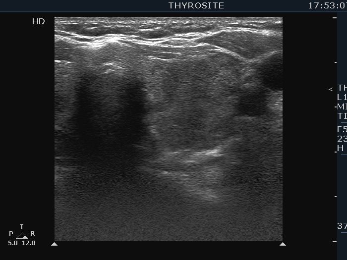
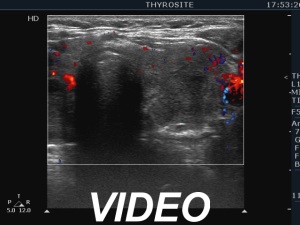
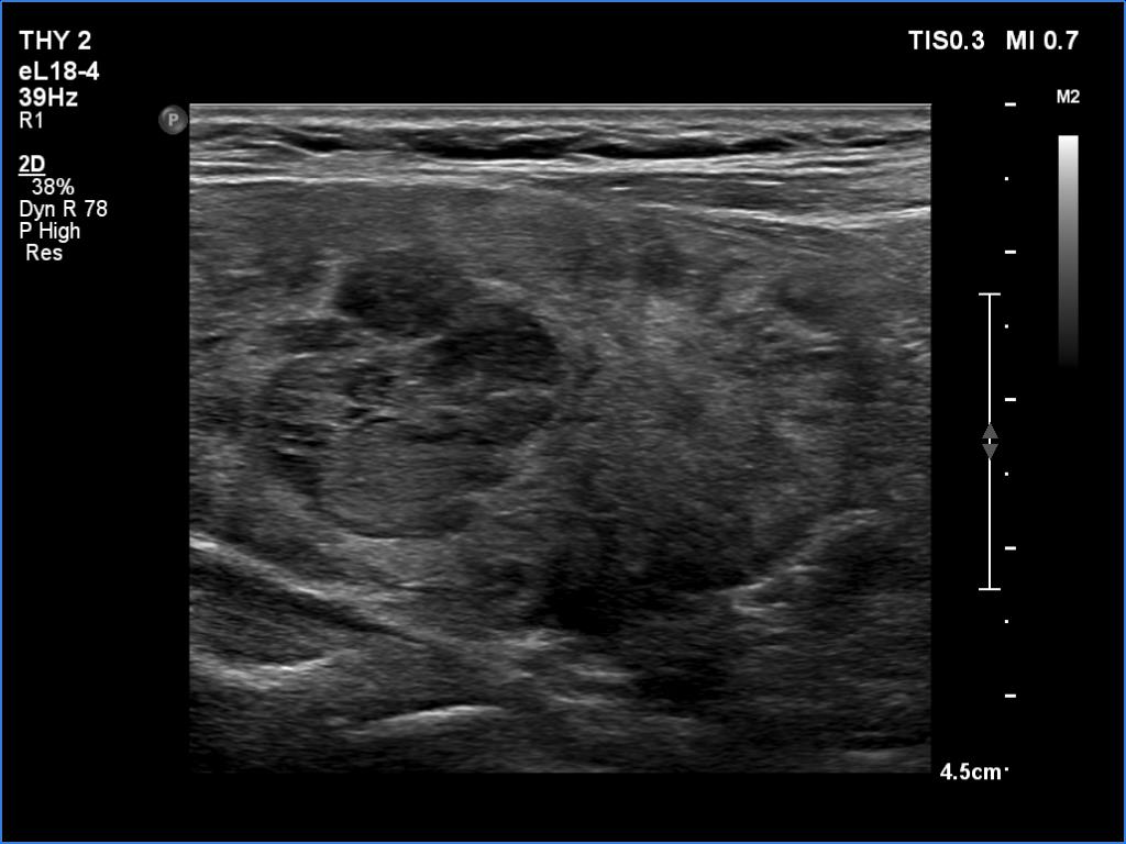
Three years after the first examination (third row of images):
Clinical presentation. The patient has ceased the thyrostatic therapy for six months. Recently, she noticed again complaints suggesting hyperthyroidism.
Palpation: unchanged.
Results of blood tests: hyperthyroidism (TSH undetectable, FT4 39.1 pM/L).
Ultrasonography. There were two changes compared with the previous examination. The thyroid has become echonormal while the lesion in the right lobe has become again hypoechoic.Suggestion: daily 20 mg methimazole. Radioiodine therapy when the FT4 level will become normal.
Comments.
- This case demonstrates the exception and not the rule. The echogenicity of a nodule almost always remains the same even over decades while in this patient the echogenicity has significantly changed two times within three years. The possible explanation is that the basic echo pattern of the extranodular thyroid and the vascularization influenced the pattern of the lesion.
- It is not evident whether the lesion in the right lobe is a single nodule with lobulated margins or a mass composed of multiple discrete lesions. In the former case the lobulation is pathological in the latter the undulation is not.