
|
|
Graves' disease - case 530
|
|


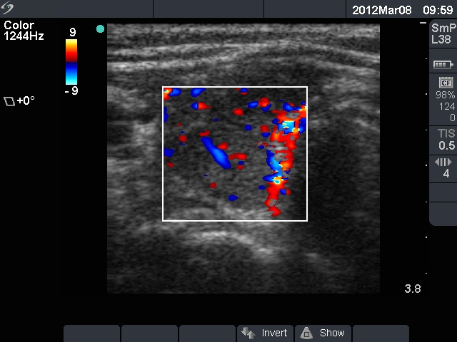
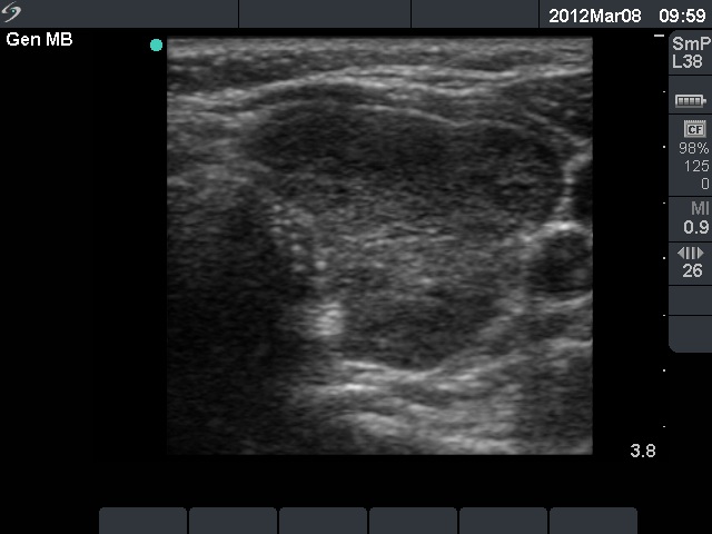
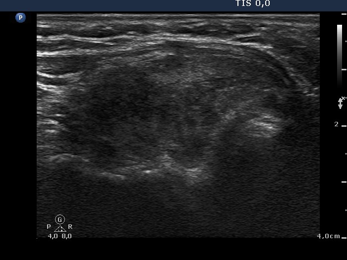
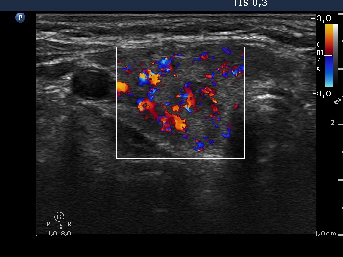
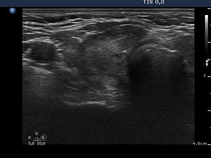

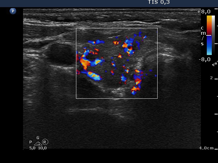
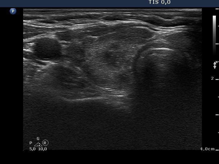


First examination (first row):
Clinical presentation: A 28-year-old woman was referred for evaluation of a recurrent hyperthyroidism. She underwent radioiodine therapy 3 years ago. She did not require replacement therapy and was euthyroid in the previous 30 months. Her original complaints recurred for two months, including 8 kg loss in weight and tachycardia.
Palpation: Both thyroids were enlarged and moderately firm.
Result of blood test: hyperthyroidism (TSH undetectable, FT4 38.6 pM/L).
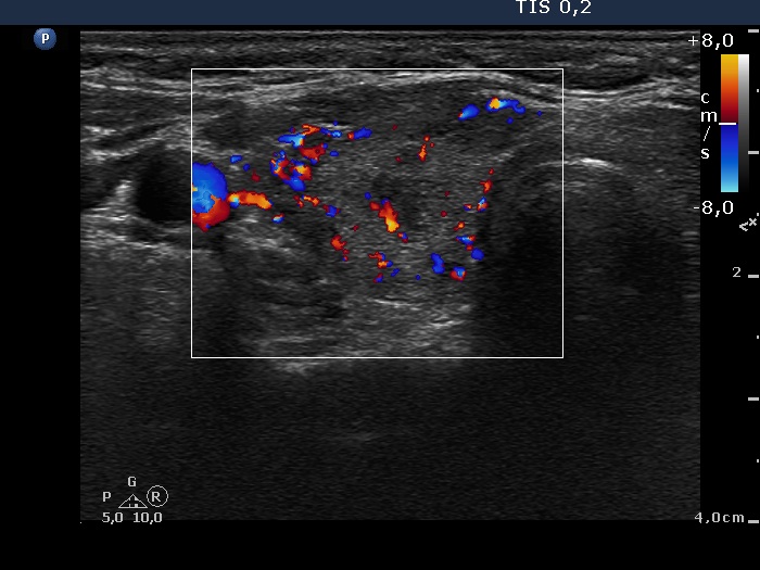
Ultrasonography: A diffusely hypoechogenic thyroid was found with multiple more hypoechogenic lesions. None of them corresponded to a nodule. The vascularization was increased.Daily 20 mg methimazole was administered and repeat radioiodine therapy was advised after reaching the euthyroid state.
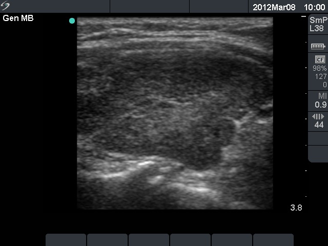
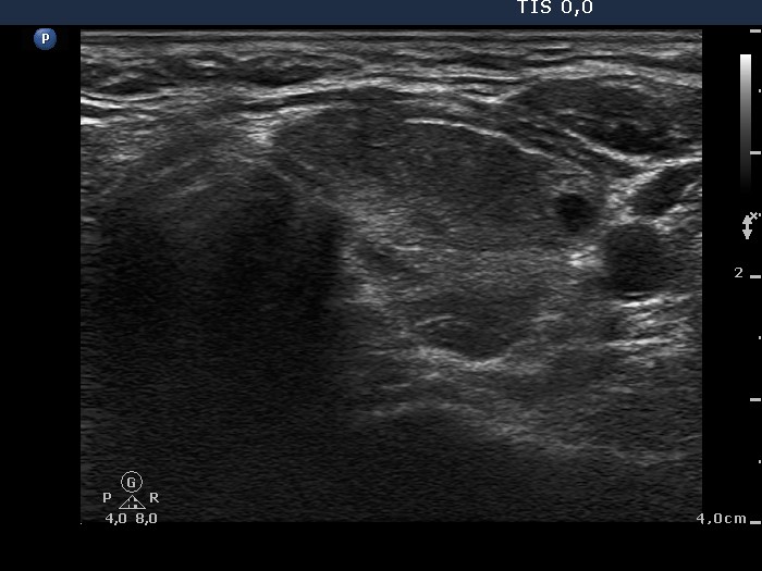
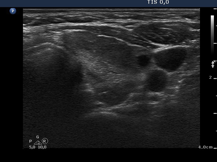
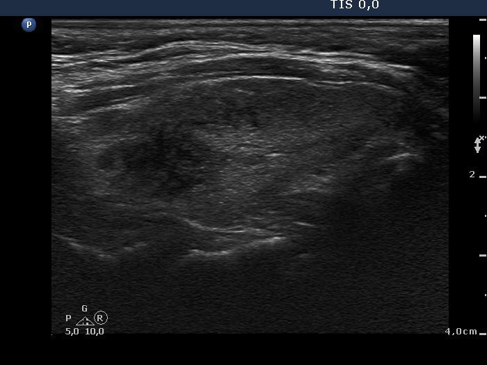
Follow-up examination 12 months later (second row):
Clinical presentation: The patient did not undergo radioiodine therapy. She was well and euthyroid on thyrostatic therapy in the previous year.
Palpation: Both thyroids were enlarged and moderately firm.
Result of blood test: subclinical hyperthyroidism on daily 10 mg methimazole (TSH undetectable, FT4 13.4 pM/L, FT3 5.05 pM/L).
Ultrasonography: The extent of hypoechogenic areas have decreased, otherwise the pattern was unchanged.We suggested again a repeat radioiodine therapy.
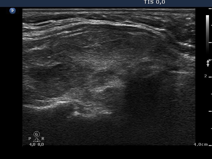
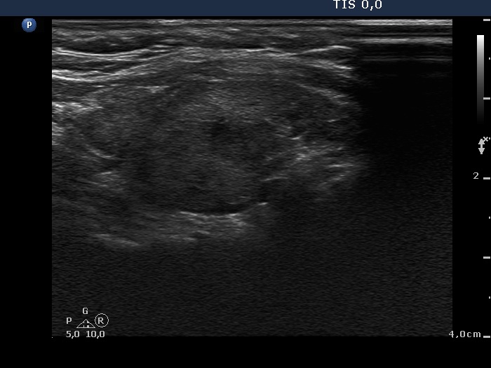
Third examination 18 months after initial examination (third row):
Clinical data: Another relapse of hyperthyroidism occurred 3 months ago. Thereafter, the dose of thyrostatic was increased.
Palpation: unchanged.
Result of blood test: subclinical hyperthyroidism (TSH 0.03 mIU/L, FT4 18.1 pM/L).
Ultrasonography: The pattern was the same.
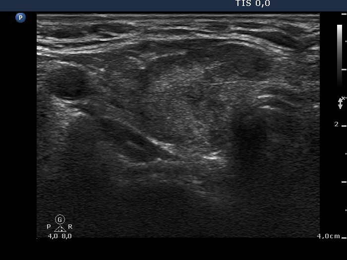
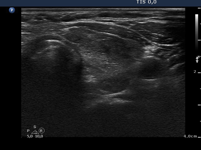
Fourth examination 21 months after initial investigation (fourth and fifth rows):
Clinical presentation: The patient requested a second opinion. Multiple nodules including a suspicious one in the right lobe were described in another institute. Scintigraphy diagnosed multiple "cold" and warm" nodules in both lobes. Surgery was advised. The surgeon asked aspiration cytology of the hypoechogenic nodule described as suspicious on ultrasonography.
Palpation: unchanged.
Results of blood tests: euthyroidism on daily 25 mg methimazole (TSH 3.78 mIU/L, FT4 10.9 pM/L).
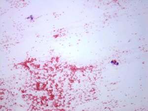
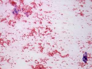
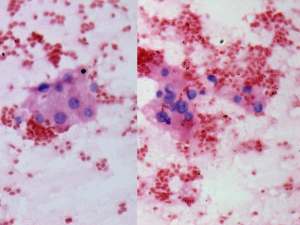
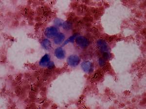
Ultrasonography: unchanged.Cytology was performed from the hypoechogenic area in the upper part of the right thyroid.
Cytological diagnosis: benign pattern corresponding to previous dysfunction and isotope therapy.
The patient underwent total thyroidectomy. Histopathology disclosed diffuse goiter corresponding to Graves' disease and focal lymphocytic thyroiditis. There were no nodules.
Comment. It is worth analyzing the echo pattern of the thyroid. The small hypoechogenic area in the right lobe changed neither in size nor is shape nor in vascularization over 11 months.