
|
|
Parathyroid lesions - case 878
|
|


Clinical data: A 61-year-old woman was referred for preoperative localization of a parathyroid adenoma. The patient has been treated for hypothyroidism for seven years. Recently, hyperparathyroidism was diagnosed on evaluation of osteoporosis. MIBI scintigraphy disclosed increased uptake according to one of the left parathyroids.
Palpation: no abnormality.
Laboratory tests: TSH 2.81 mIU/L on daily 75 microgram levothyroxine. Serum-parathormone 117.2 pg/mL (normal value 12-88), serum calcium 2.97 mM/L, phosphorus 0.60 mM/L.
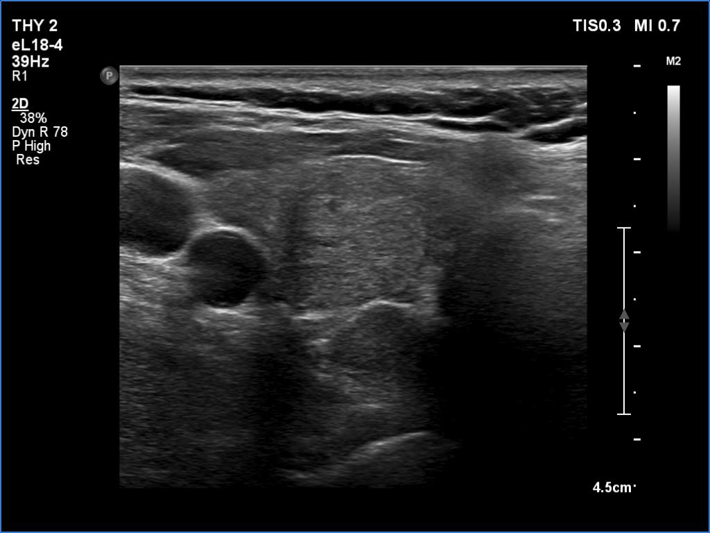
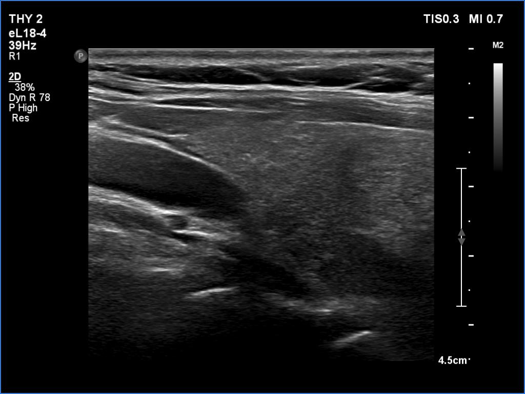
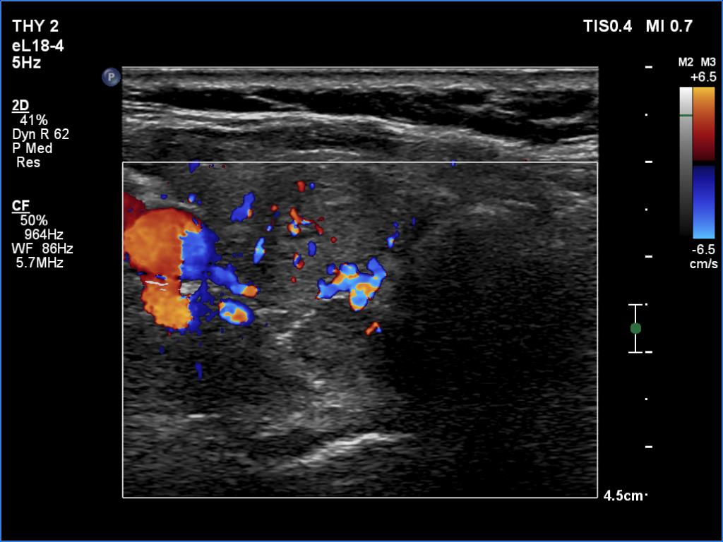
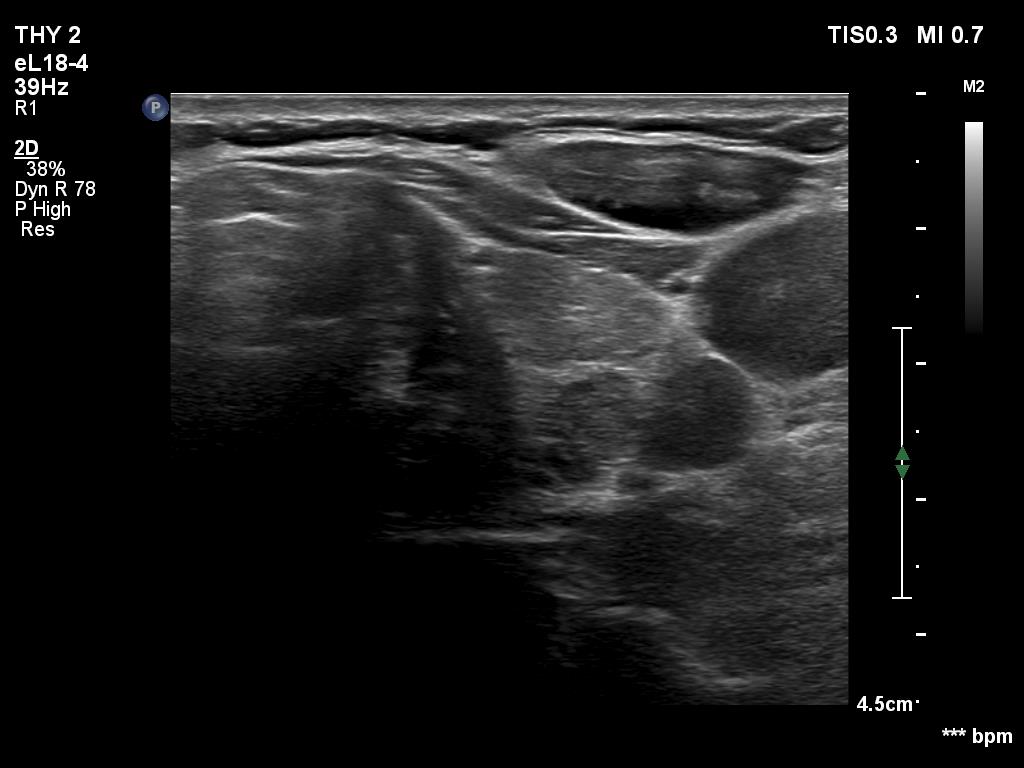
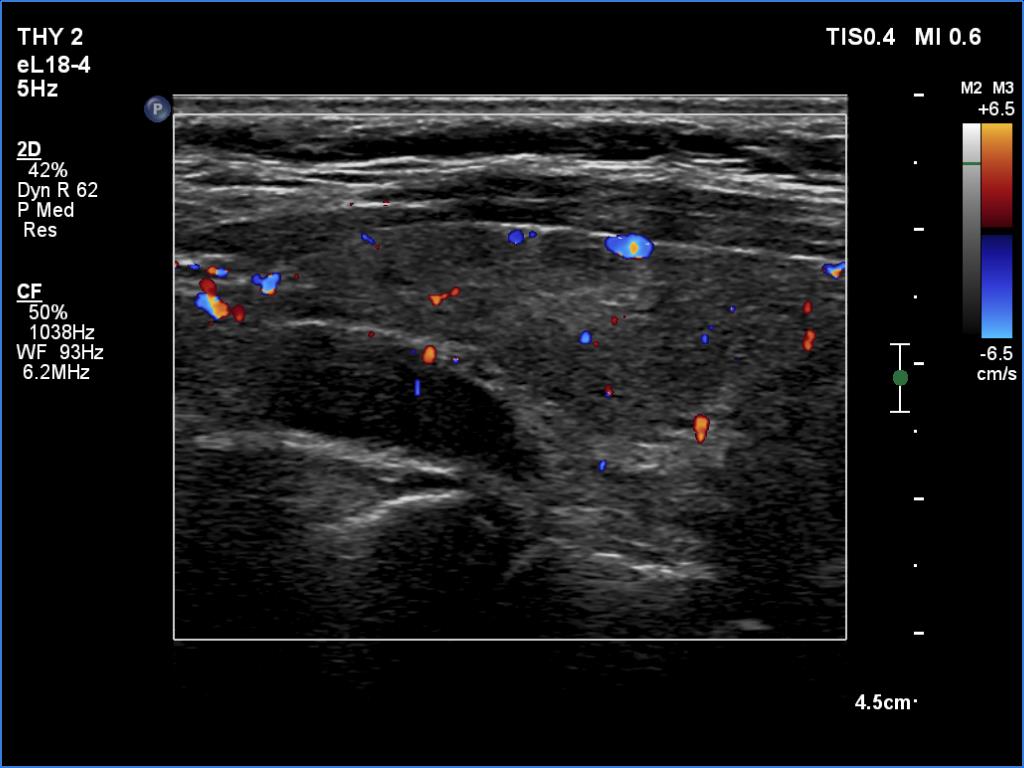
Ultrasonography. The thyroid was moderately hypoechoic. There were two discrete lesions in the right lobe while a hypoechoic mass was found dorsal to the middle third of the left lobe.
During surgery, intraoperative parathormone measurement disclosed elevated levels according to the left upper parathyroid which has been removed. Histopathology: parathyroid adenoma.