
|
|
Ethanol sclerotherapy: thyroid cysts - Case 11
|
|


First session (first row of images):
Clinical presentation: A 50-year-old man requested an examination. He had a cystic nodule which was drained two times in the previous year. The size of the nodule was 25x18x37 mm, 25x15x34 mm and 23x16x35 mm (6.74 mL), thirteen months, 6 months, and 2 months prior to this visit. After both aspirations, the maximal diameter of the lesion became less than 16 mm. At this time, the patient felt a recurrence of the cyst and came to the planned sclerotherapy.
Palpation: a moderately firm nodule in the left lobe.
Functional state: euthyroidism with TSH 1.96 mIU/L.
Ultrasonography: The thyroid was echonormal and inhomogeneous. There was a central-type cyst with a minimally hypoechogenic solid part. The lesion displayed signs of perinodular blood flow.
Three sessions of sclerotherapies were applied in 3 weeks. At the first occasion, 2 mL serous fluid was aspirated and 2.5 mL ethanol was given. Later 1.2 and 1 mL ethanol were administered, at the 2nd and 3rd session, respectively.
Three months after the therapy (second row of images):
Clinical presentation: The patient had no complaints and found the lesion having been disappeared.
Palpation: no abnormality.
Functional state: euthyroidism with TSH 1.99 mIU/L.
Ultrasound: The nodule has significantly decreased in size. We noticed the presence of intranodular hyperechogenic granules and signs of intranodular blood flow.
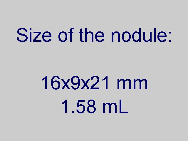
Three years after the therapy (third row of images):
Clinical presentation: The patient had no complaints.
Palpation: no abnormality.
Functional state: euthyroidism with TSH 2.73 mIU/L.
Ultrasound: The nodule became a bit larger. We observed more intranodular hyperechogenic granules and the degree of intranodular blood flow has increased.
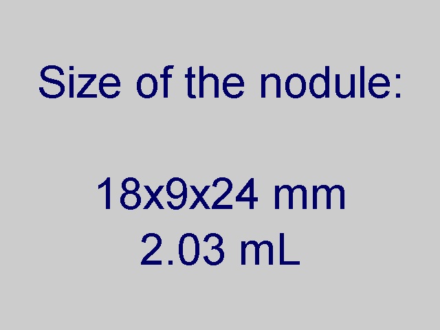
Four years after the therapy (fourth row of images):
Clinical presentation: the patient had no complaints.
Palpation: The left lobe was suspicious harboring a nodule.
Functional state: euthyroidism with TSH 3.86 mIU/L.
Ultrasonography. The nodule became greatee and displayed perinodular blood flow.
Six years after the therapy (fifth row of images):
Clinical presentation: The patient had no complaints.
Palpation: a firm nodule in the left lobe.
Functional state: euthyroidism with TSH 2.89 mIU/L.
Ultrasonography. The volume of the lesion has increased further. The volume of the lesion was 78% of pretreatment volume.
Comments.
-
There is a statistical issue to which size we have to compare the follow-up findings, to the original size or the size at the first session. This nodule has refilled repeatedly. At the time of the first session, we did not wait to a full refilling therefore the size at the first session is significantly lower than two months before this time.
-
The appearance of intranodular hyperechogenic granules including microcalcification-like figures is a frequent finding in PEI-treated nodules and might cause concern if we are not aware of this possibility.