
|
|
The issue of large goiters - case 120
|
|

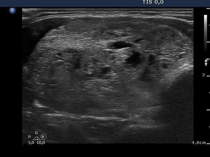


Clinical data: A 46-year-old woman came to a follow-up examination. She had multinodular goiter known for years. She noticed that her right thyroid had increased over the last two years. Sometimes she had difficulties in swallowing.
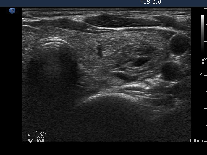
Palpation: The right lobe was enlarged, multiple nodules were palpated in both lobes.
Functional state: euthyroidism (TSH 0.38 mIU/L, FT4 12.0 pM/L).
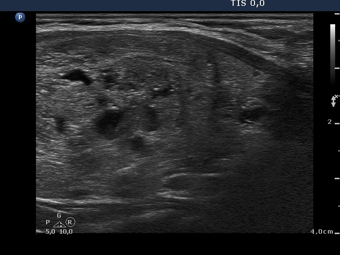
Ultrasonography: Both lobes contained multiple nodules with different echogenicities. The lower pole of the right thyroid could not be visualized in supine position before swallowing, while during swallowing it came into sight.
A combined sonographic-cytological diagnosis of benign lesion was given.
Surgery was performed.
Histopathology disclosed benign hyperplastic nodular goiter.
Comments.
-
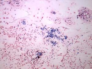
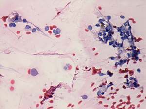
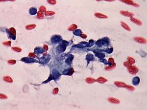
The cytological pattern presents signs of atypia. Atypical follicular cells were in abortive microfollicles. Although there were no signs of papillary or a Hürthle-cell tumor, the pattern may correspond to atypia of unknown significance and on the cytological pattern itself we cannot exclude the possibility of follicular carcinoma. On the other hand, the sonographic pattern excluded a follicular tumor.
-
There are three degrees of substernal spread:
-
Mild degree: the lower pole of the thyroid cannot be visualized uprightly but can be in supine position with extended neck. It is very common in elderly patients (first of all of in men) even in the absence of thyroid enlargement.
-
Moderate degree: the lower pole cannot be visualized in supine position with extended neck before swallowing, but comes insight while swallowing. The goiter demonstrated in this case belongs to this category.
-
Severe degree: the lower pole cannot be visualized in supine position with extended neck even while swallowing.
-