
|
|
The issue of large goiters - case 1736
|
|


Clinical data: A 74-year-old woman was referred for ultrasound examination. The patient got radioiodine therapy 21 years ago. A relapsing hyperthyroidism was first noticed 5 years after the treatment. I met the patient 8 years ago when she was euthyroid on methimazole therapy, and the size of the nodule in the right lobe was 42x34x51 mm. Because the goiter caused no complaints and the patient tolerated thyrostatic therapy well, she refused to get a repeat radioiodine therapy.
Palpation: a non-firm large nodule in the right lobe.
Laboratory tests: TSH 0.19 mIU/L, FT4 17.2 pM/L on daily 15 mg methimazole.
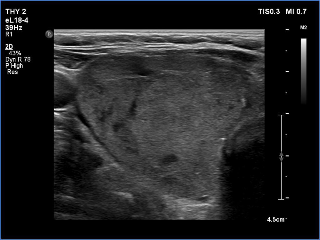
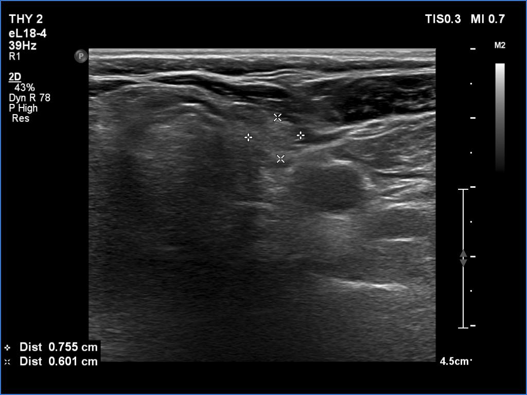
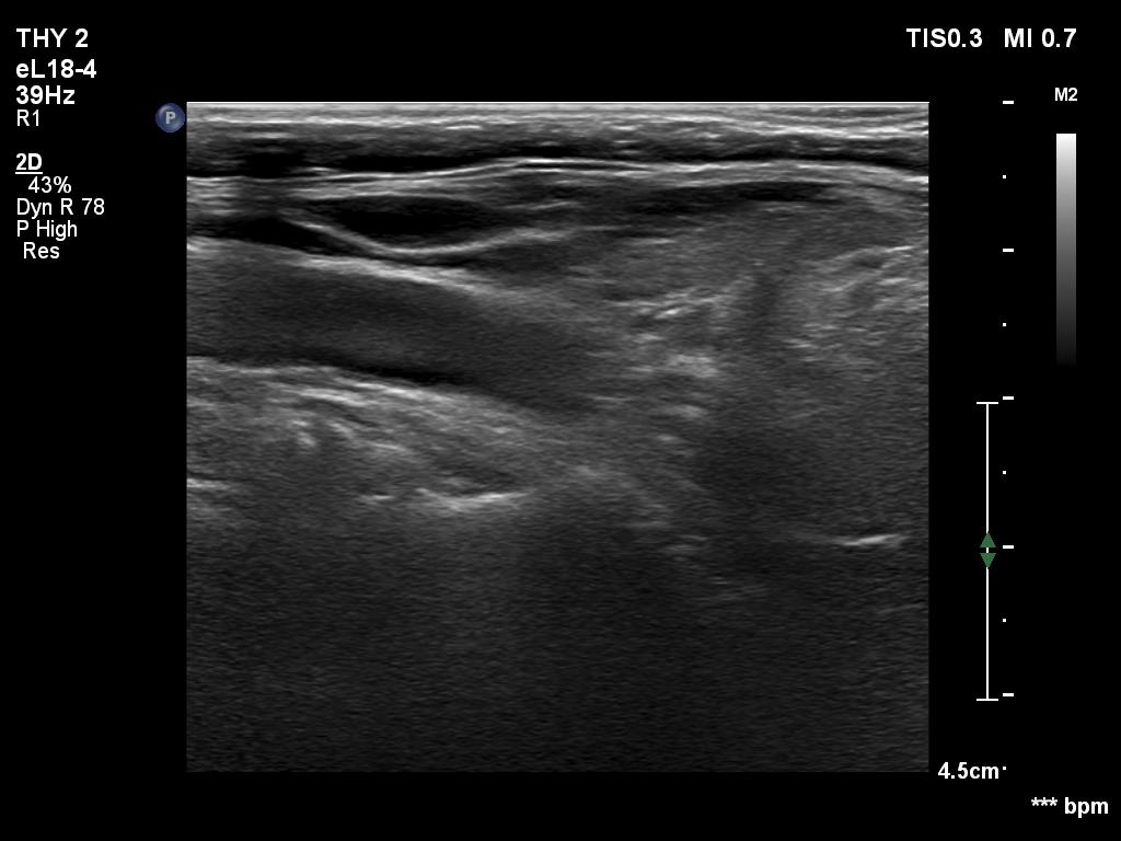
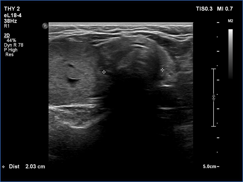
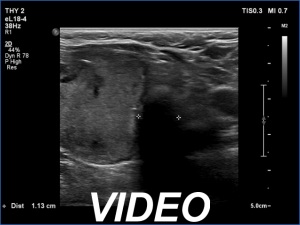
Ultrasonography. The right lobe was enlarged and had an echonormal nodule. The lesion shed both intranodular and perinodular vascularity. The dimensions of the nodule were 42x34x53 mm The left lobe was very small in size. Compared with the upper third of the thyroid, the trachea was narrowed to half in diameter at the lower third of the thyroid.
Comment.
-
A long-standing autonomously functioning adenoma causes a continuous shrinkage of the contralateral lobe.
-
The combined perinodular and intranodular vascularity is typical of an autonomously functioning adenoma.