
|
|
Case 364
|

|


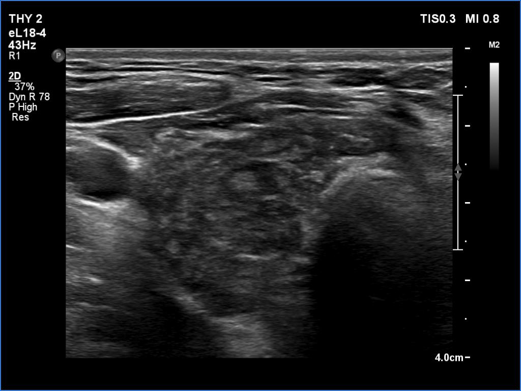
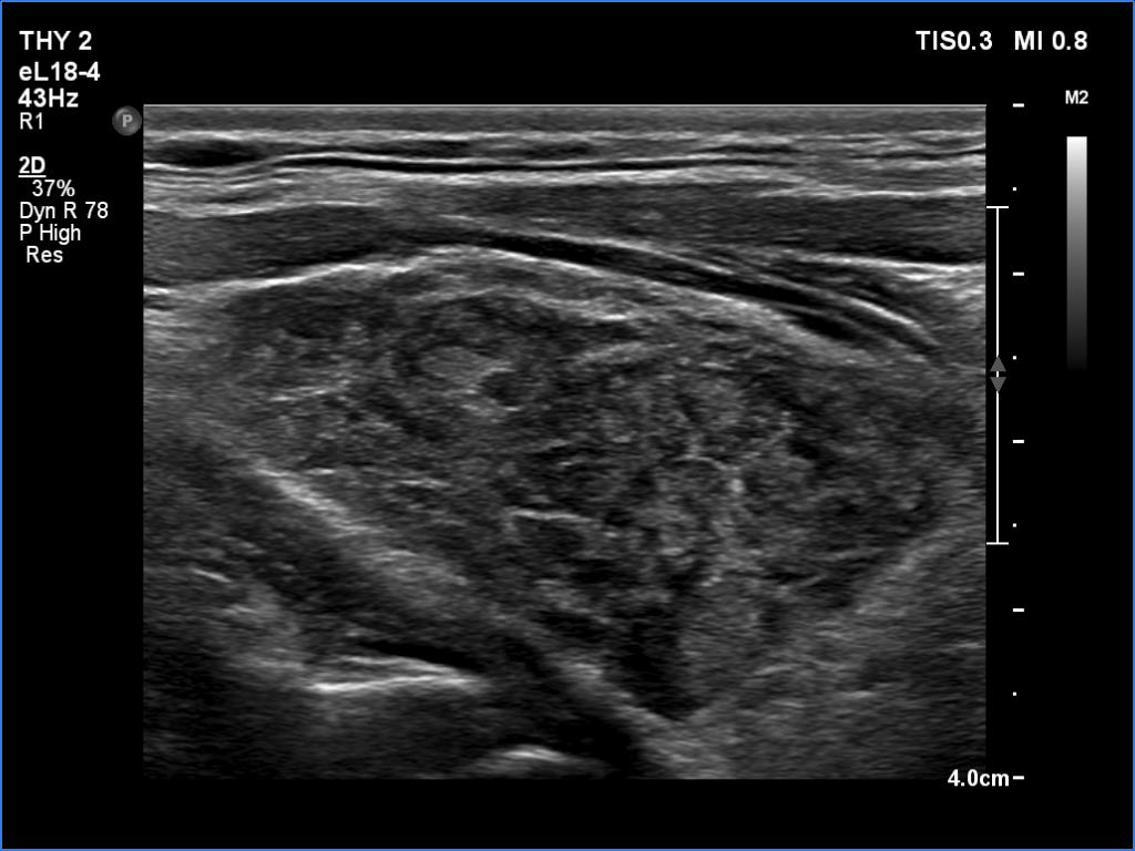
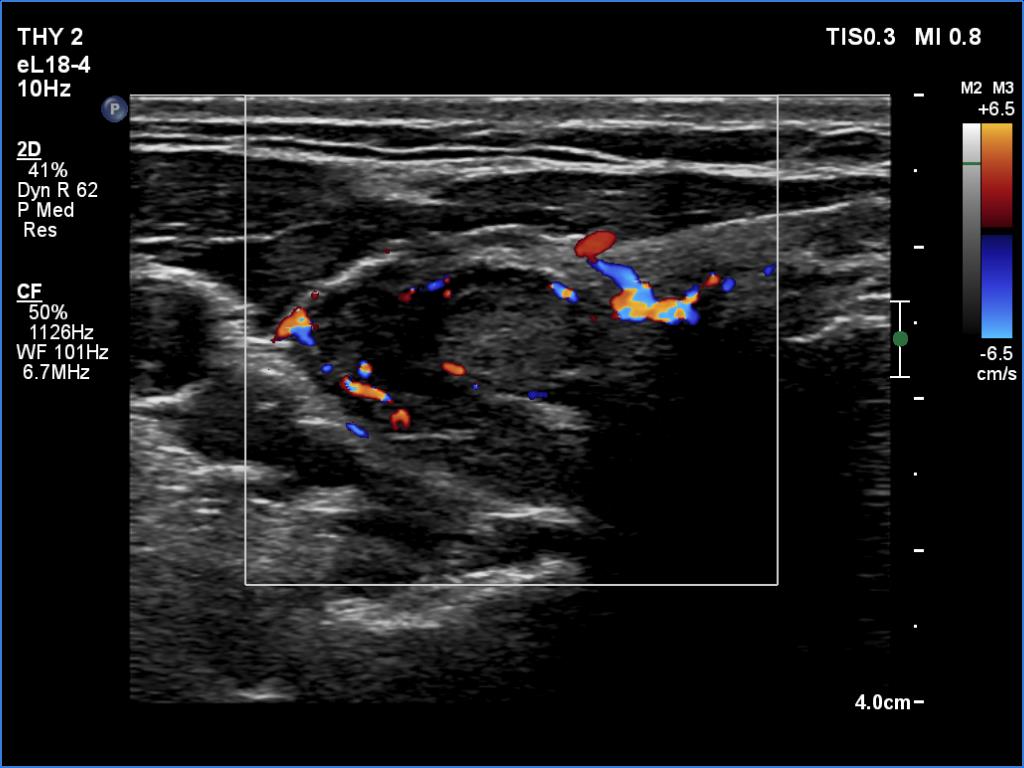
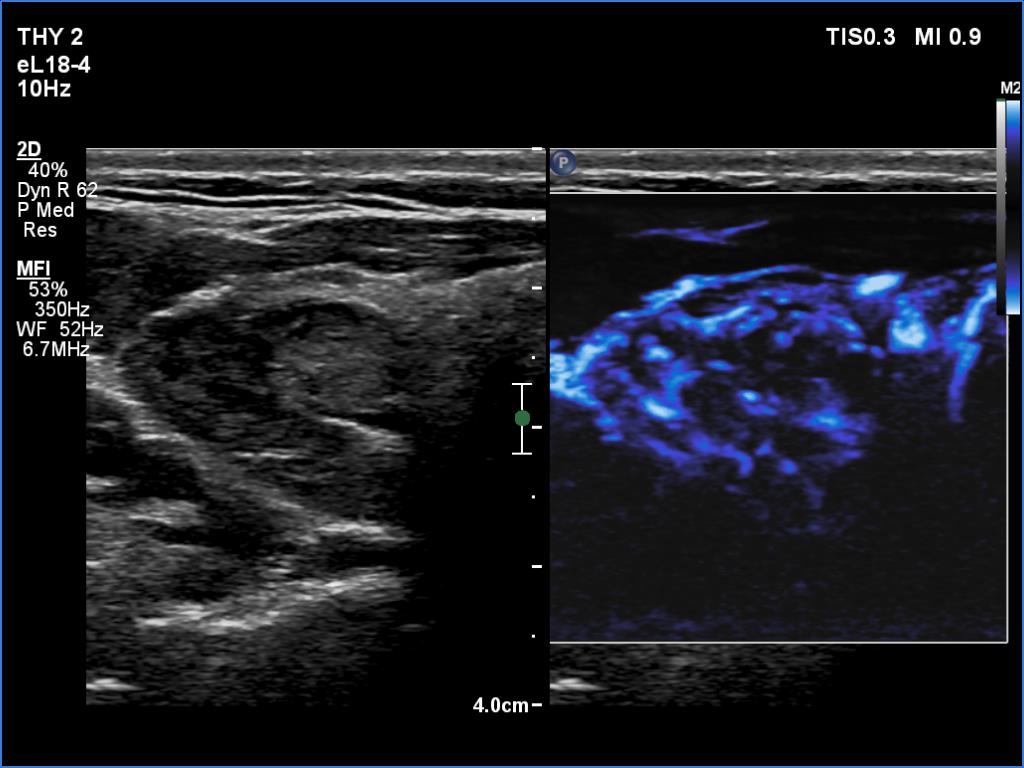


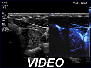
Clinical data: A 52-year-old man requested a second opinion. He had been treated for hypothyroidism for 9 years. Recently, on thyroid ultrasound a suspicious nodule with microcalcifications was described in the right lobe.
Palpation: no abnormality.
Laboratory tests: TSH 8.03 mIU/L on daily 75 microgram levothyroxine.
Ultrasound. Both lobes were hypoechogenic. There were numerous less hypoechoic discrete areas corresponding to those parts of the gland which were not or less influenced by the underlying thyroiditis. The largest of these lesions was in the right lobe. It had both echogenic lines and granules, a pattern corresponding not to microcalcifications but connective tissue.
Aspiration cytology from the hyperechogenic lesion resulted in Hashimoto's thyroiditis.
Suggestion: to increase the dose of levothyroxine to daily 100 micrograms. Repeat ultrasound in 5 years.
Comment. The discrete lesion is more probably the largest of the intact part of the thyroid than a true nodule. Note that there are numerous, discrete areas with similar echogenicity also in other parts of the thyroid. The lesion in question is also infiltrated by thyroiditis.


