
The operated thyroid - case 337 (ultrasonographic picture 6b)
|
|

|
|
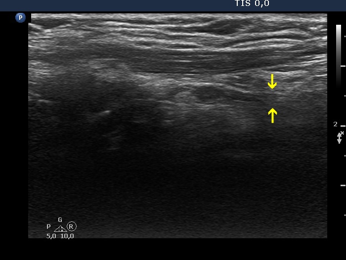
Left lobe, longitudinal scan. The hypoechoic mass has upper and lower (yellow arrows) tails which means that this does not correspond to thyroid tissue but to muscle fiber.