
100 consecutive cases of papillary cancer - case 066 (ultrasonographic picture 7)
|
|

|
|
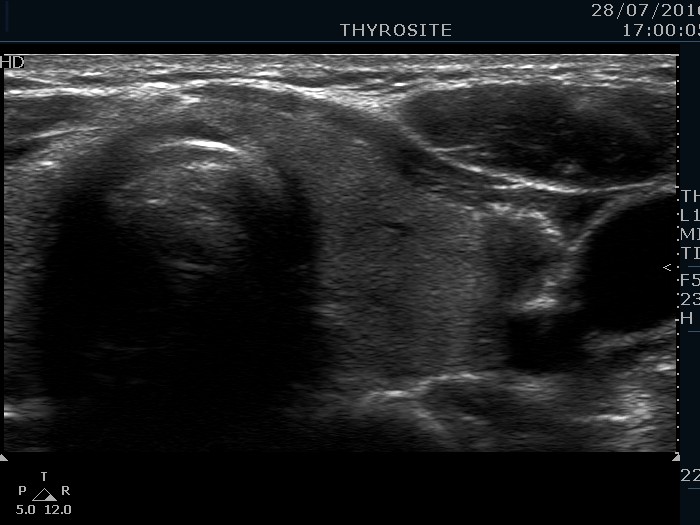
Left lobe, trasnverse scan. This lobe has small hypoechoic areas including a hypoechoic nodule in the lateral part which shows taller-than-wide shape.