
100 consecutive cases of papillary cancer - case 066 (ultrasonographic picture 8)
|
|

|
|
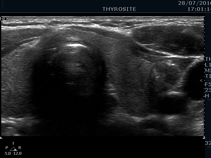
Lower part of the left lobe, transverse scan. There is a nodule in the lateral part of the lobe. The lesion has echogenic granules. Only the most brightest is suspicious being microcalcification.