
100 consecutive cases of papillary cancer - case 085 (ultrasonographic picture 4)
|
|

|
|
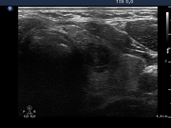
Left lobe, transverse scan. There is a hypoechogenic lesion in the central part of the lobe. It contained small hyperechogenic punctate granules resembling microcalcification. See the video record.