De Quervain's thyroiditis - Case 11 |
|
 |
 |
 |
 |
 |
 |
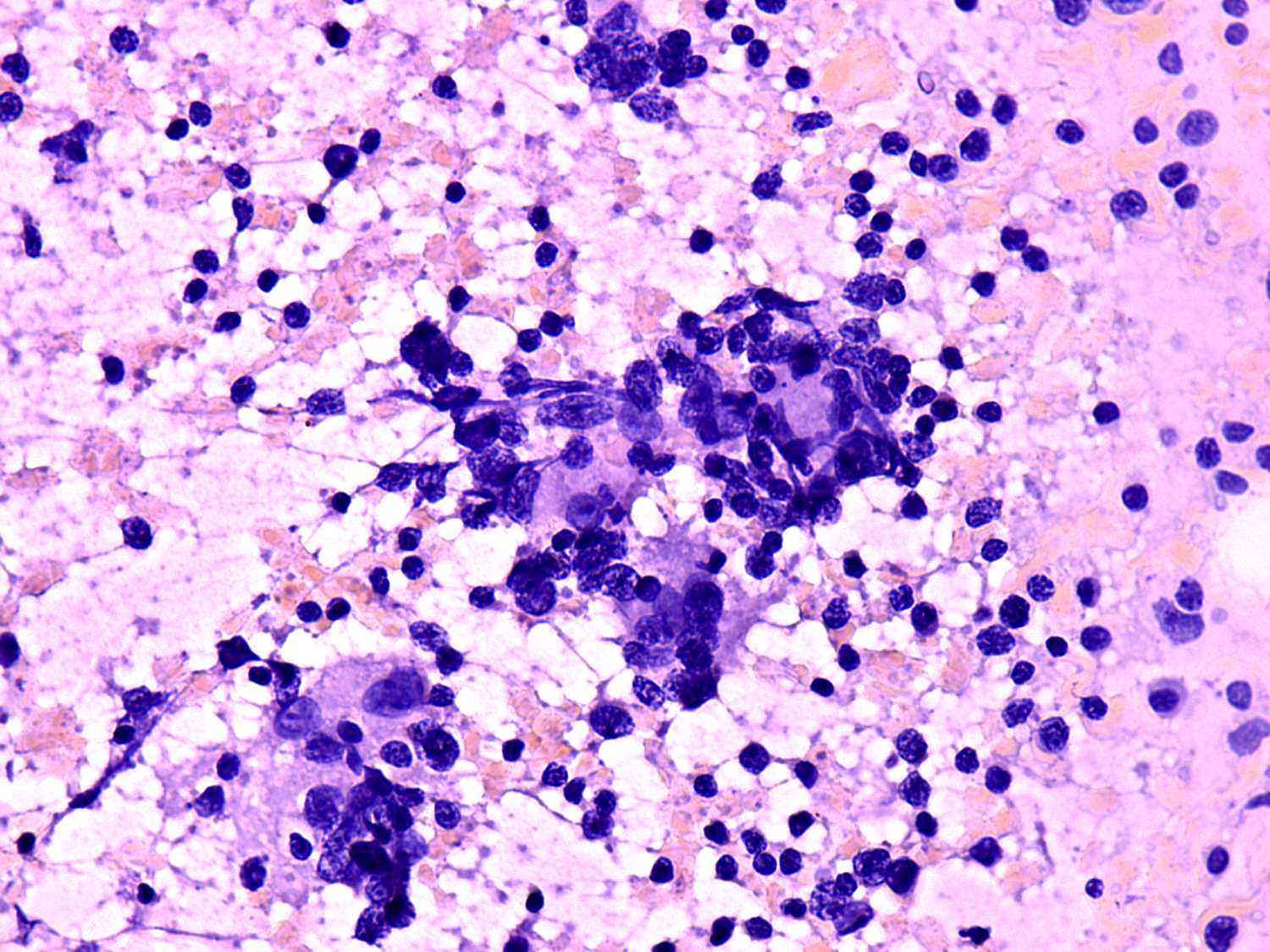
The right images present most characteristic sign of de Quervain's
thyroiditis, i.e. multinucleated cells composed of elongated epitheloid
cells. |
| |
|
 |
 |
 |
 |
 |
 |
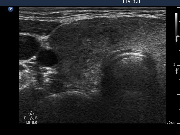
The sonographic presentation seems to be very similar but in fact the
two cases differ in small but significant properties. The
thyroids are basically hypothyroid and there are small echonormal areas
present in both cases. However, in the de Quervain patient the
echonomal areas
are mainly outside the hypoechogenic part and in the
Hashimoto's case within the hypoechogenic part. The cases
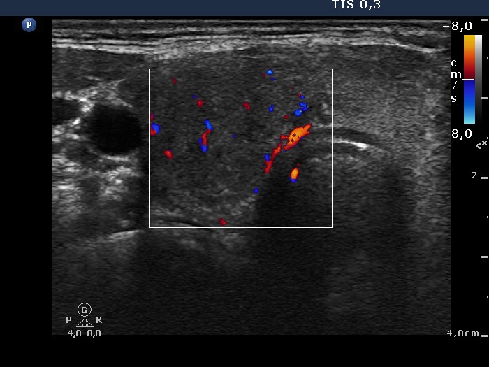
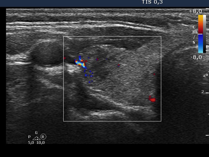
differ characteristically in vascularization, as well. The De Quervain
case presents the typical decreased vascular pattern. A decreased
vascularization itself does not exclude the presence of a Hashimoto's
thyroiditis, while conversely an increased vascularization practically
excludes the possibility of de Quervain's thyroiditis. |
|
And last but not least the clinical data:
|
|
Three weeks history of
subfebrility
and thyroid pain
|
History
|
A patient treated for hypothyoidism
without neck complaints |
|
Hard, painful
|
Palpation
|
Firm, painless |
|
Not performed
|
aTPO
|
428 U/ml |
|
70 mm/H
|
ESR
|
Normal |
| |
|
|
|
| |
|
| |


