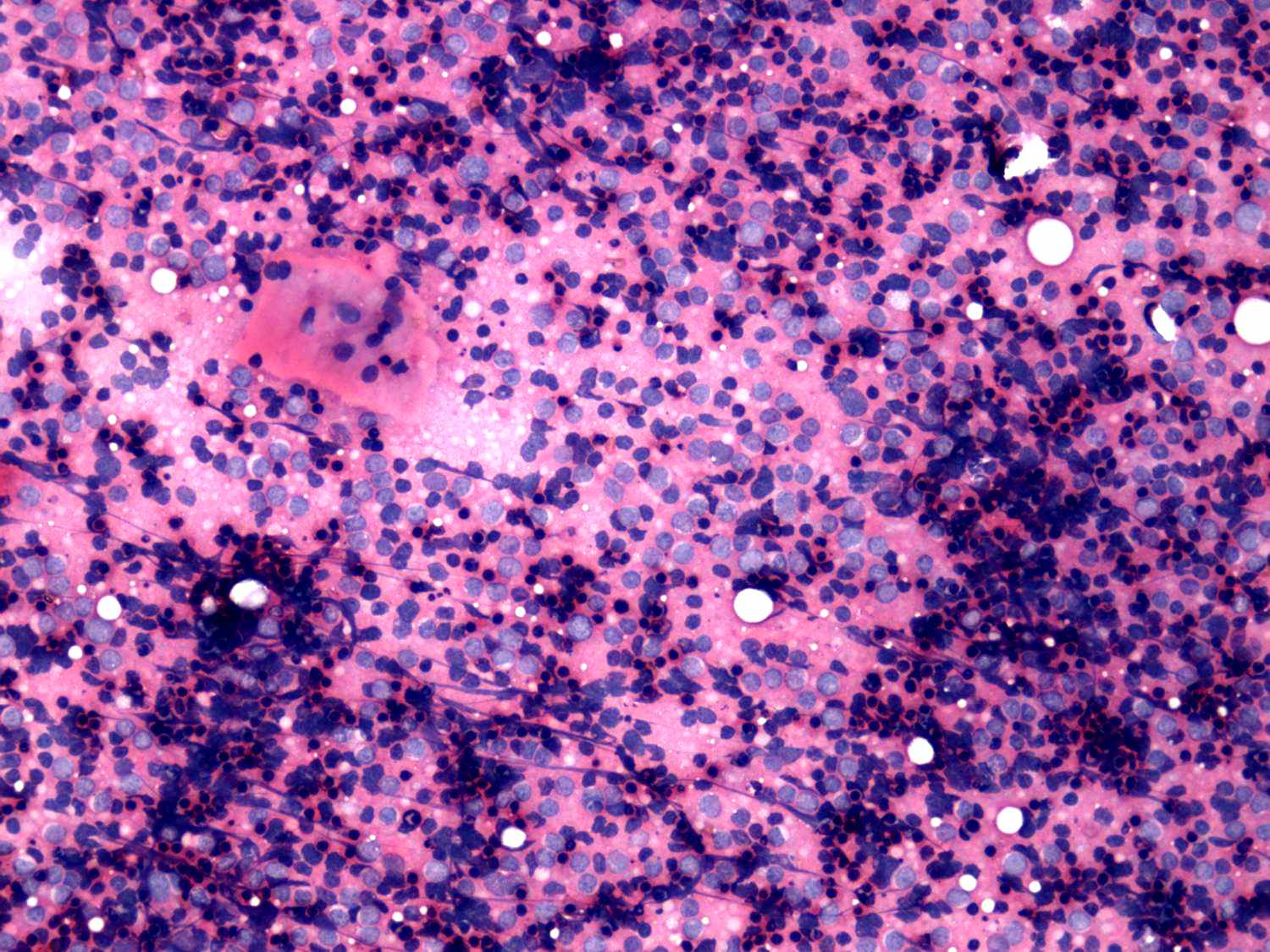
The cytological pattern is identical in these cases: heterogeneous
lymphoid cell population predominate the smear with small clusters of
oxyphilic cells.
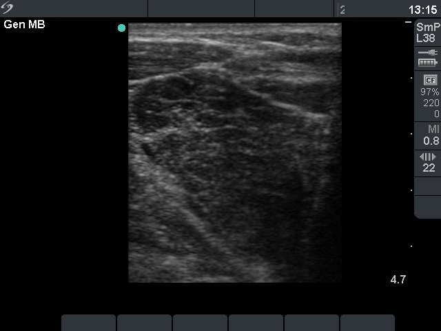
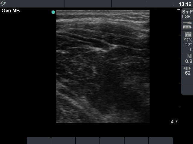
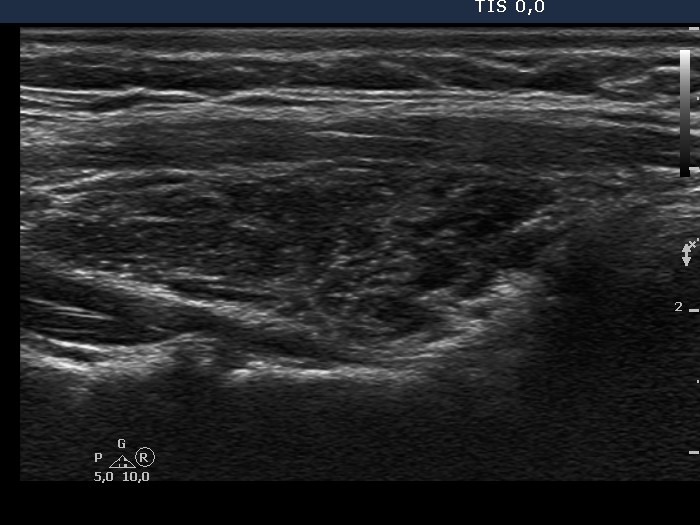
The image cannot reflect the reality: the patient presented with an
extremely enlarged thyroid evolved over 2 months. Only a part of the
right lobe can be demonstrated. The whole lobe is hypoechogenic and
contains connective tissue, as well.
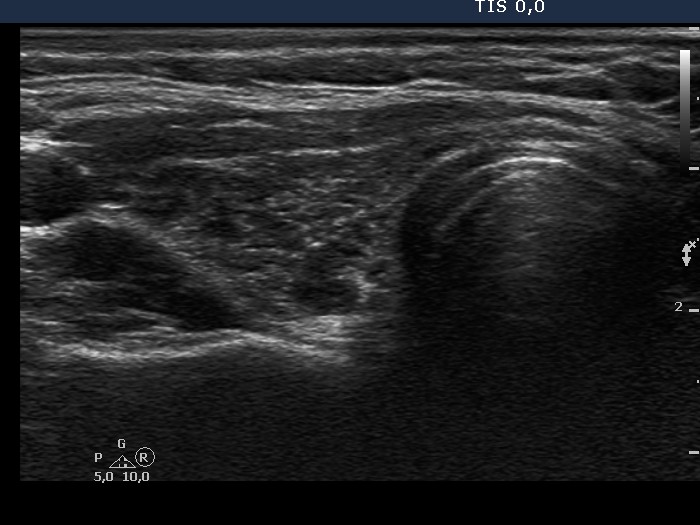
The thyroid was not enlarged. There are several hypoechogenic lesions
within an echonormal background.
Only anaplastic carcinoma and MALT-lymphoma are involved in the
differential diagnostics of such rapidly increasing huge painless
thyroid mass, while such cytological pattern may be occur only in
Hashimoto's thyroiditis and MALT-lymphoma. The situation is simple: a
combined clinical-sonographic-cytologicaal diagnosis of suspicion of
MALT-lymphoma can be given. As a rule, a definitive diagnosis of a
lymphoma mustn't give cytologically.