
|
|
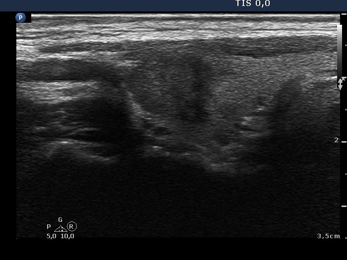
The borders of the nodule - conp 020
|
|


Clinical presentation: A 43-year-old woman was referred for follow-up examination. We met the patient for the first time 13 years ago when a hypoechogenic lesion was found on ultrasound examination. The dimensions of the lesion were 7x7x9 mm, width x depth x length, respectively. A repeat ultrasound was suggested in 5 years.
Palpation: no abnormality.
Functional state: euthyroidism (TSH 0.67 mIU/L, anti-TPO 8 U/mL).
Ultrasonography. The thyroid was echonormal. There was a hypoechogenic lesion in the upper, ventro-lateral part of the right lobe. The lesion presented partly lobular, partly blurred surface. The dimensions of the nodule were 9x12x13 mm, width x depth x length, respectively, it means that the volume of the lesion increased by more than 200% in the past 13 years.
Cytology resulted in papillary carcinoma.
Histopathology disclosed papillary carcinoma in the right lobe.
Comment. More than 50% of the borders of the nodule are ill-defined which means pathological degree of blur. Moreover, the lesion displays lobulated margins, as well.