Selected topics - intranodular hyperechogenic figures - Table 8 (large). Hyperechogenic granules in the solid part of cystic nodules |
||
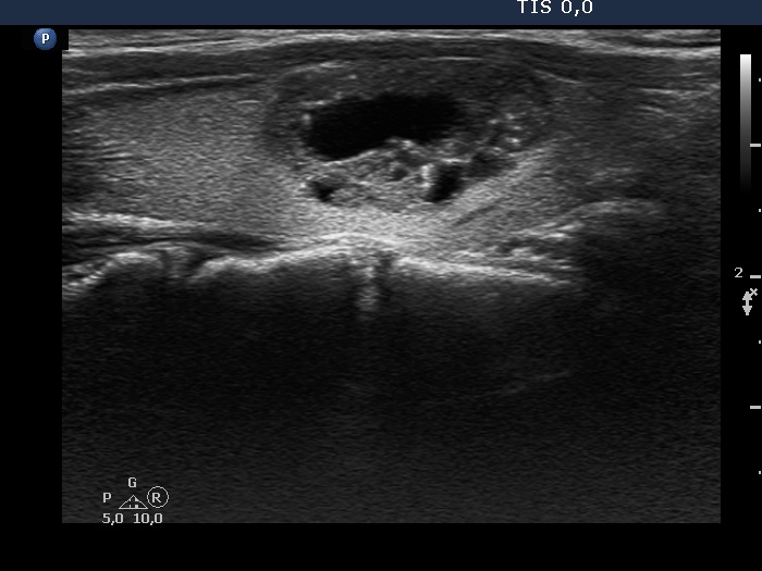
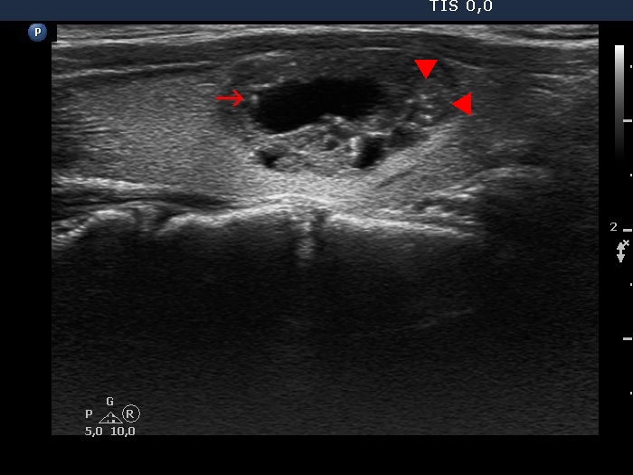
It is evident that a hyperechogenic figure in the cystic area has no oncological significance. The issue is the interpretation of granules within the solid part of a mixed lesion and within solid nodules. A colloid crystal might appear in the latter, as well and the presentation is less typical because of optical reasons: the typical tail is much better visualized in a fluid than in a solid area.
Another problem is caused by the posterior back wall enhancement in microcystic lesions which can cause broad spectrum of optical artifacts including granules similar to punctate echogenic foci (microcalcifications).
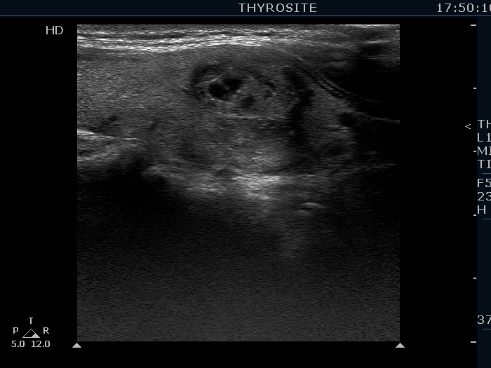
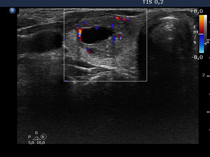
Benign cystic-colloid goiter (cytological diagnosis) |
|
 |
 |
|
|
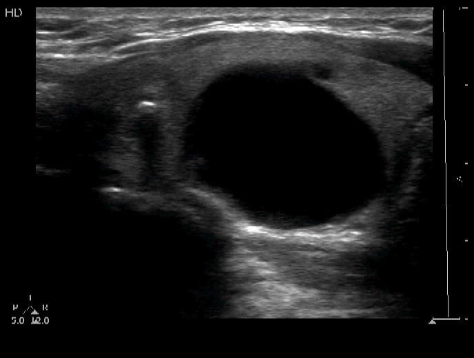
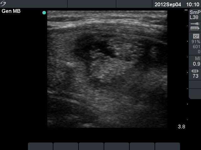
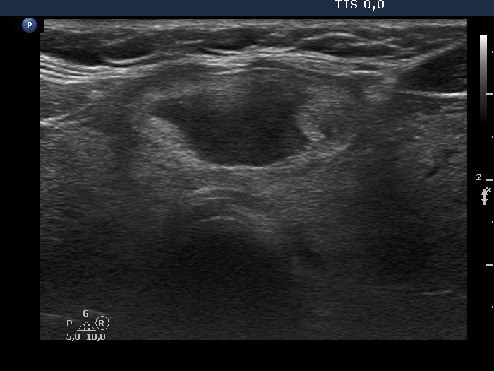
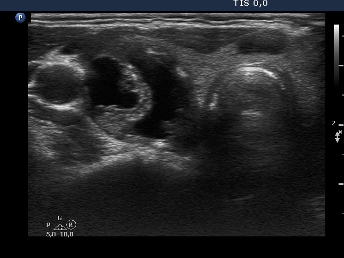
Follicular adenoma (histological diagnosis) |
|
Before aspiration |
|
 |
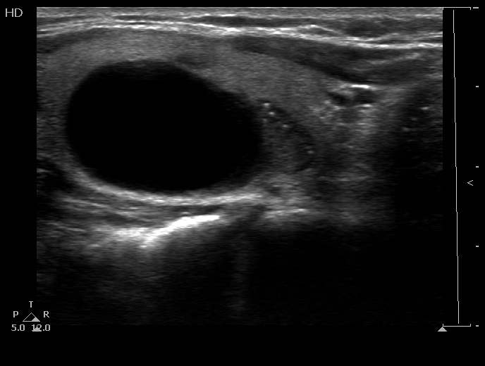 |
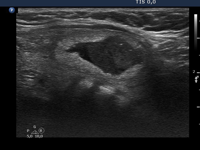
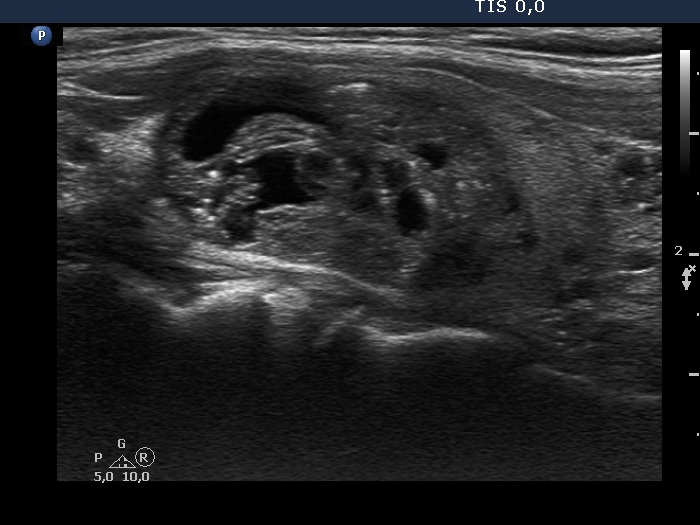
After aspiration 6.5 ml cystic fluid |
|
 |
 |
|
|
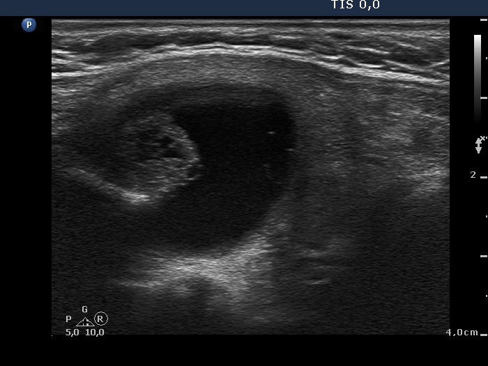
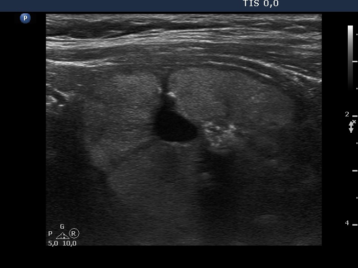
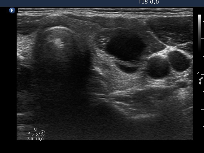
Benign cystic-colloid goiter (cytological diagnosis) - case 1473 |
|
 |
 |
 |
 |
|
|
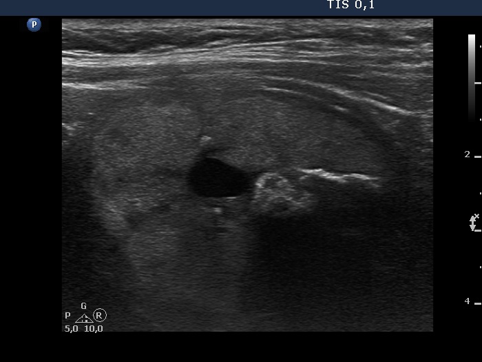
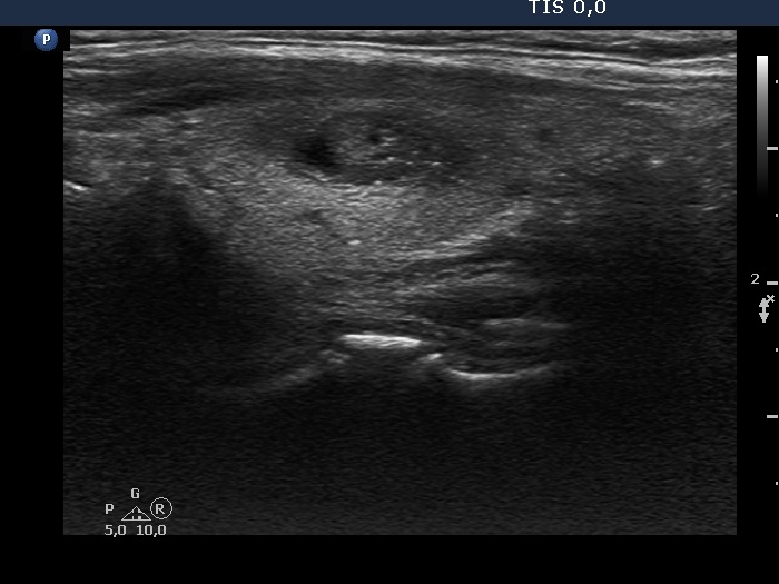
Benign cystic degeneration (cytological diagnosis) - case 1669 |
|
 |
 |
 |
 |
|
|
Papillary carcinoma (histological diagnosis) - case 853 |
|
 |
 |
|
|
Benign cystic degeneration (cytological diagnosis) - case 808 |
|
Before aspiration |
|
 |
 |
After aspiration of 5 mL cystic fluid |
|
 |
 |
|
|
Benign colloid goiter (cytological diagnosis) |
|
 |
 |
|
|
Benign cystic-colloid goiter (cytological diagnosis) |
|
 |
 |
|
|
Benign cystic-colloid goiter (cytological diagnosis) |
|
 |
 |
|
|