
|
|
Papillary carcinoma - Case 37.
|
|


Clinical data: A 43-year-old woman was referred for an evaluation of a newly discovered nodule.
Palpation: A nodule was palpable in the right lobe.
Functional state: euthyroidism (TSH-level 1.92 mIU/L).
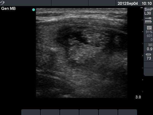
Ultrasonography revealed a mixed nodule in the right lobe. The solid part of the nodule contained microcalcification. There was a small hypoechogenic lesion in the ventral part of the left lobe. It contained both microcalcifications and coarse calcifications.
FNA was performed from both lesions. A small amount of thick brown fluid was gained from the right nodule. There were only macrophages on the smear.
Cytological diagnosis: not diagnostic and benign colloid goiter, right and left nodule, respectively.
Surgery was advised because of the sonographic presentation of the nodule.
Intraoperative imprint smears are demonstrated in case history.
Histopathology: a solitary focus of oxyphilic variant of papillary cancer with the maximal diameter of 6 mm in the right thyroid. Benign, hyperplastic nodules were found in both lobes.