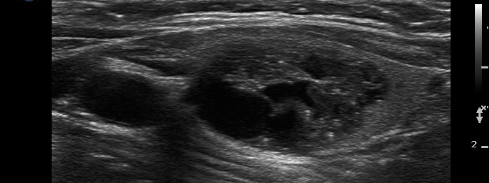
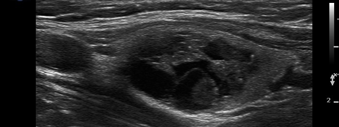
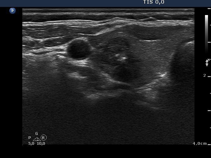
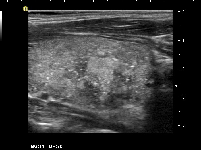
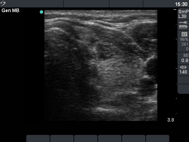
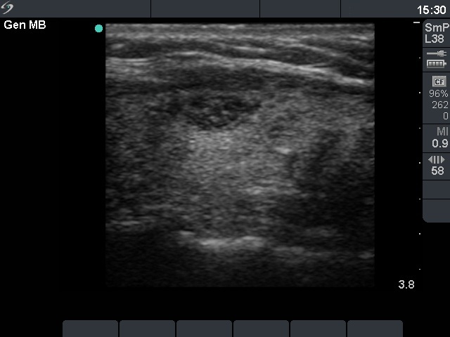
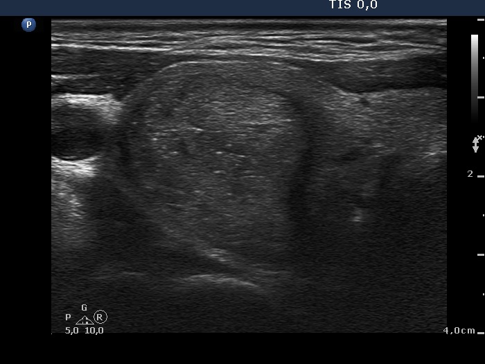
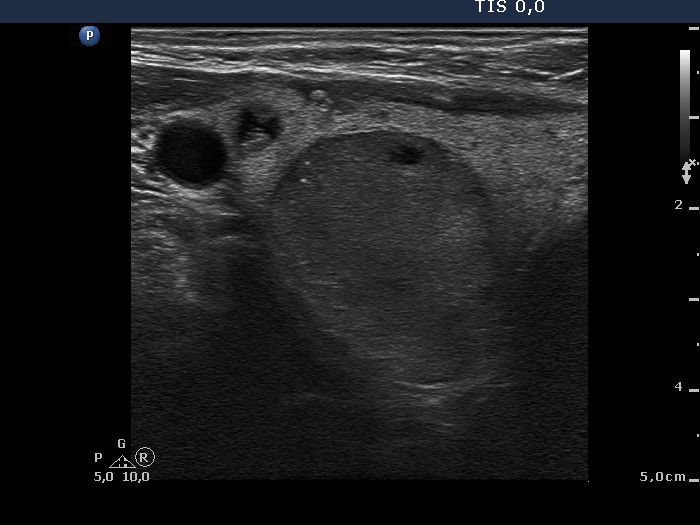
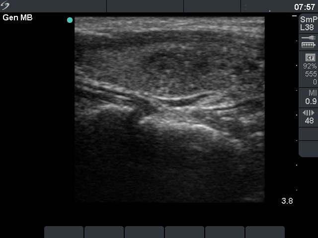
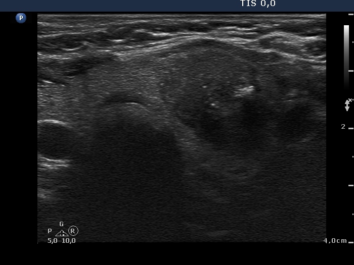
Benign hyperplastic nodule (histological diagnosis) - case 2 |
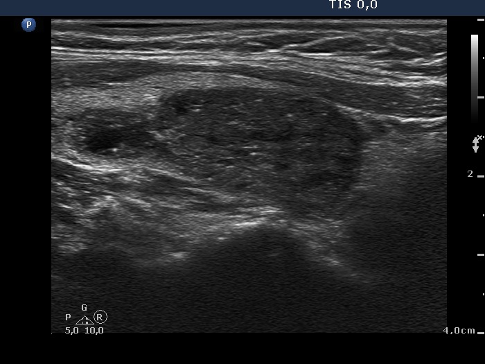
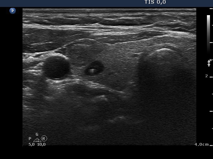
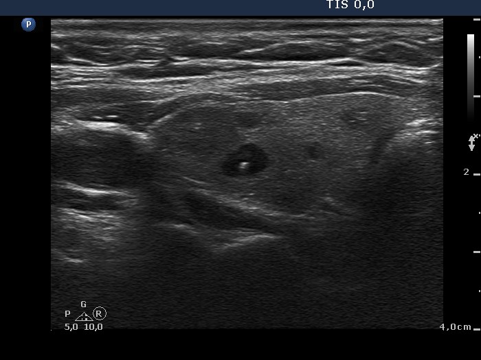
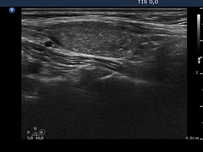
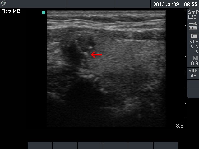
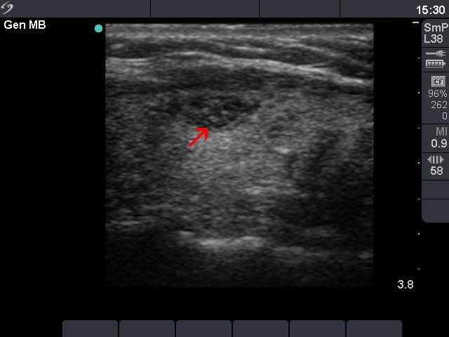
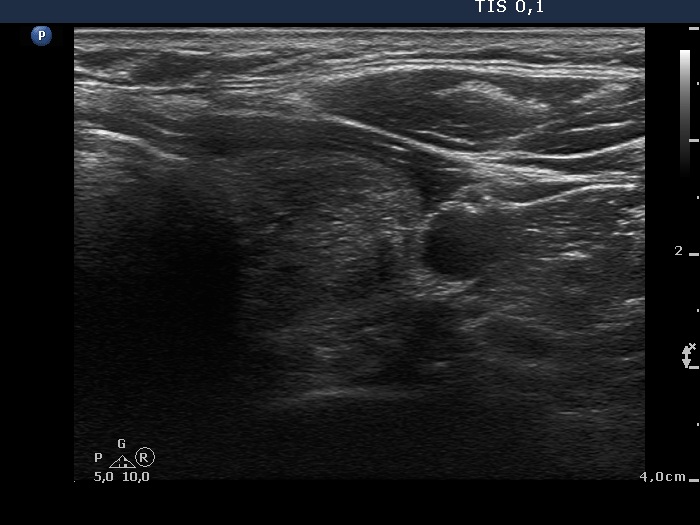
Hashimoto's thyroiditis without any nodule (histological diagnosis) - case 1520 |
 |
 |
 |
 |
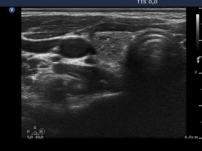
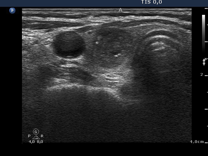
The bright granules lack the dorsal narrowing tail and there are no coexisting similarly bright lines, therefore these granules correspond to punctate echogenic foci.
|
The coexistence of tiny punctate granules and similarly bright lines is the hallmark of connective tissue. There is a brighter and relatively large granule in the ventral small lesion in the upper image which might be either a punctate echogenic focus or a sign of fibrotic changes.
|
| |
|
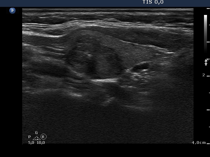
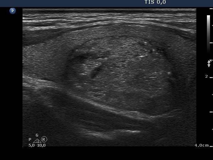
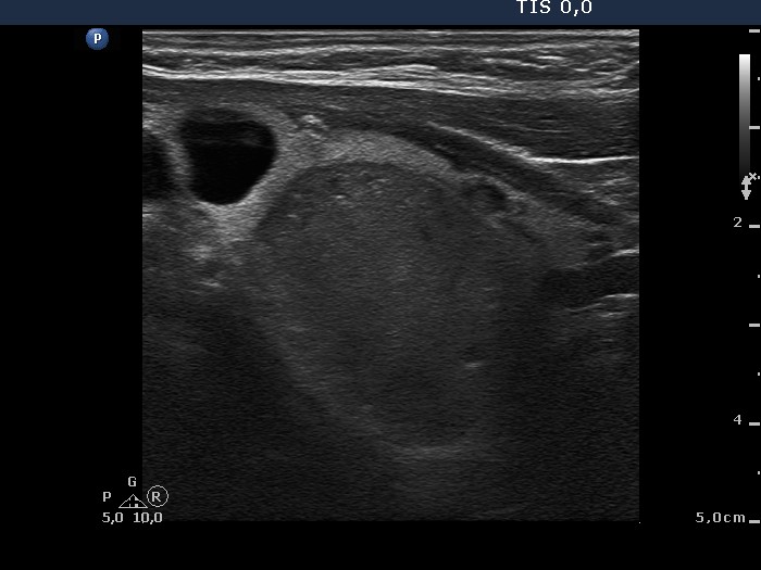
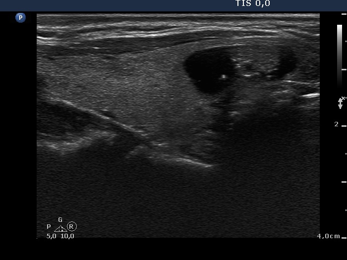
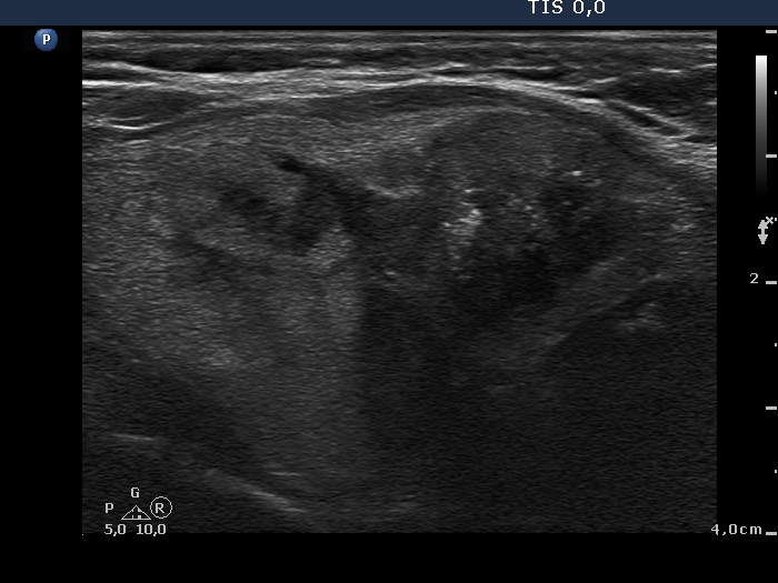
Benign hyperplastic nodules (histological diagnosis) - case cons 024
|
 |
 |
 |
 |
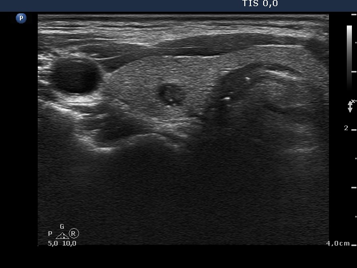
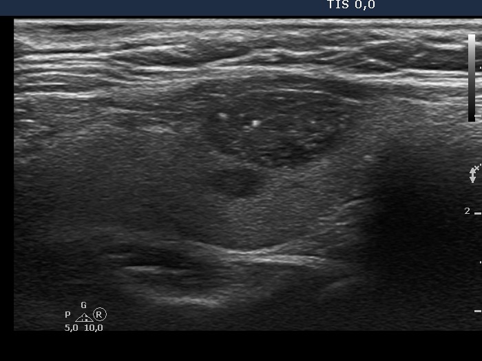
The larger lesion has a solitary bright figure which shape stands for a comet-tail artifact.
|
There are pale lines and granules within the nodule - these figures correspond to a connective tissue. The three granules are probably also presentations of a connective tissue.
|
| |
|
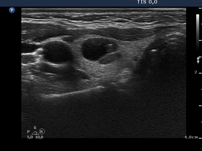
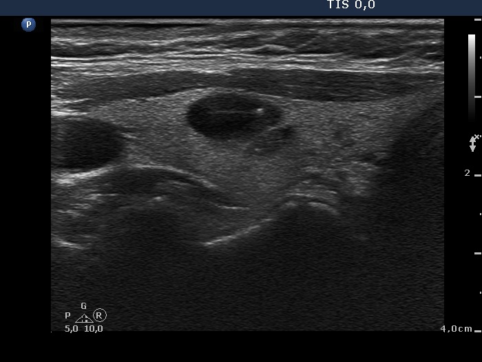
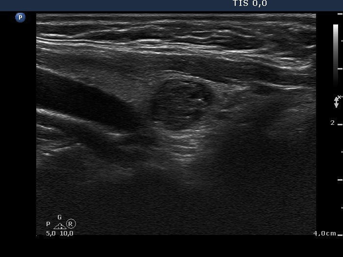
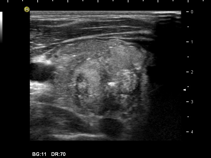
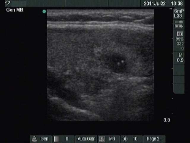
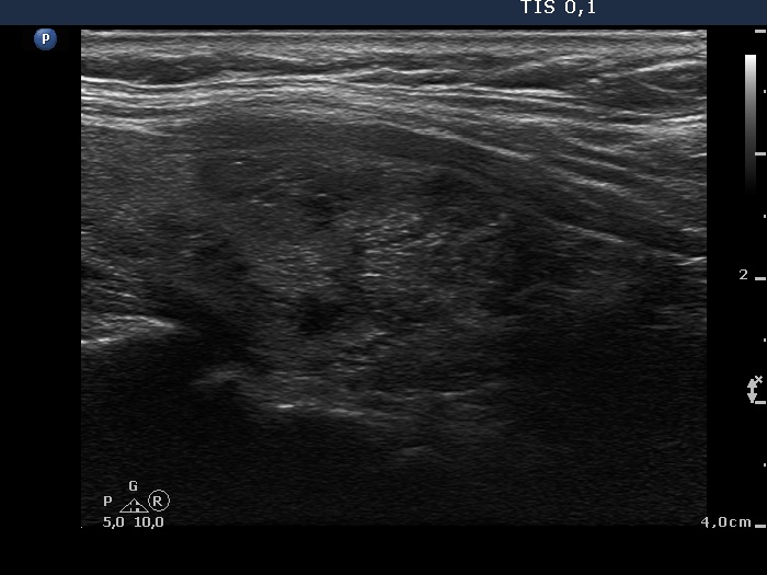
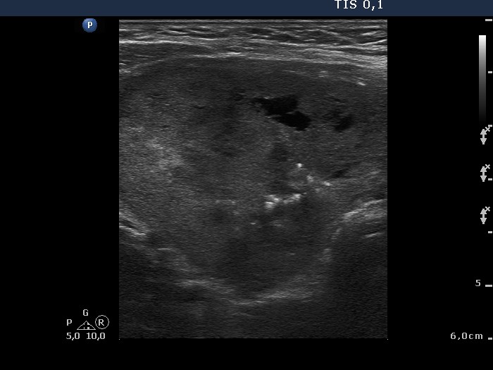
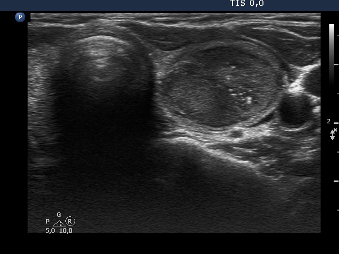
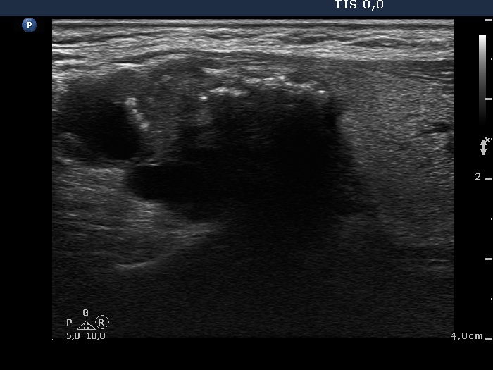
Benign cystic-colloid goiter (cytological diagnosis) - case 123 |
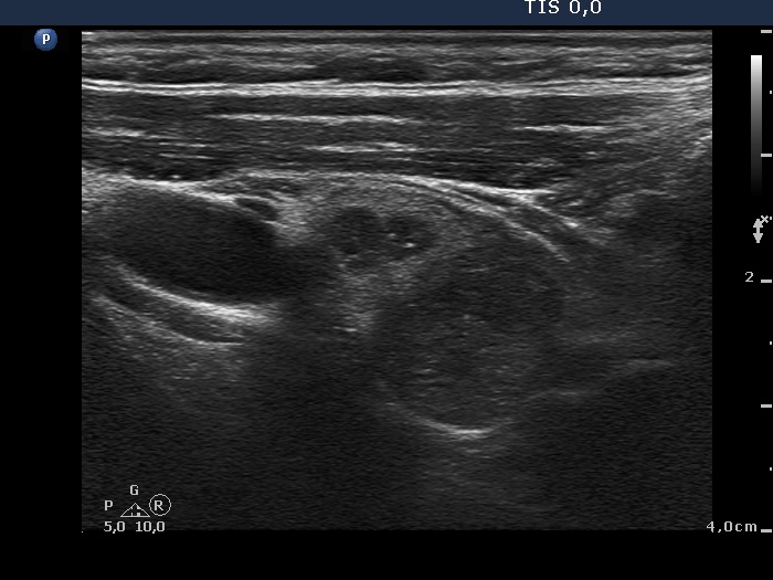
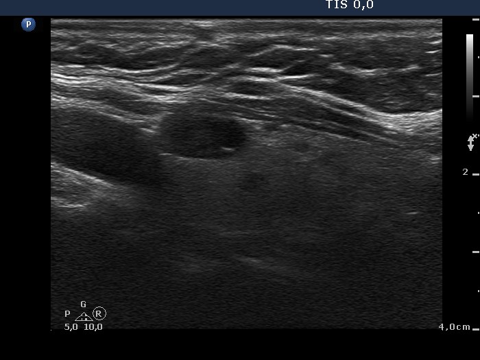
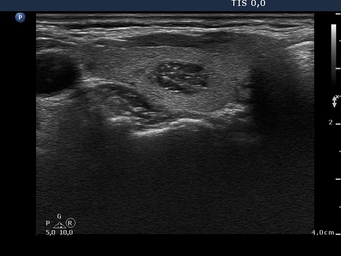
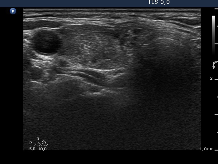
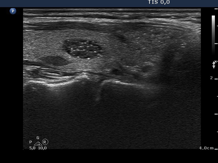
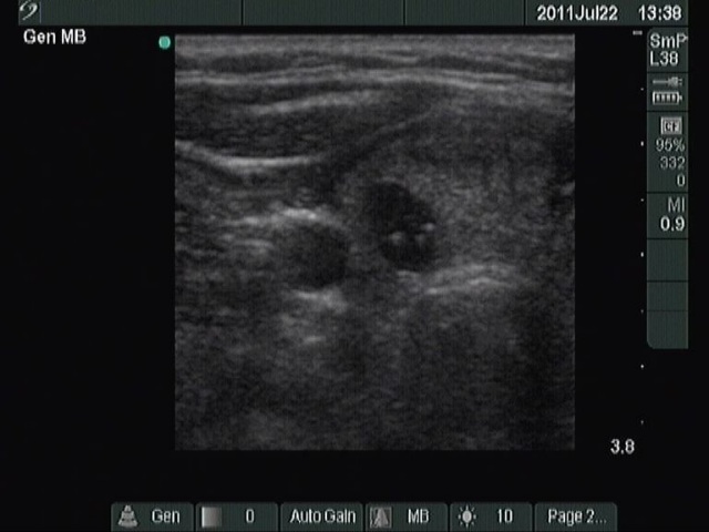
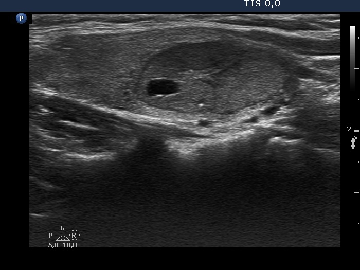
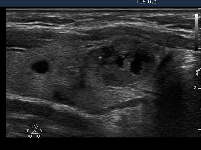
Intrathyroidal parathyroid adenoma (histological diagnosis) - case 1399 |
 |
 |
 |
 |
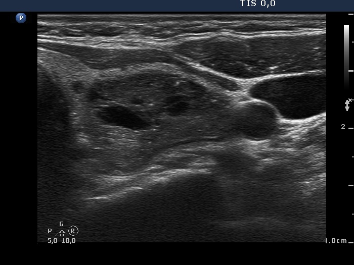
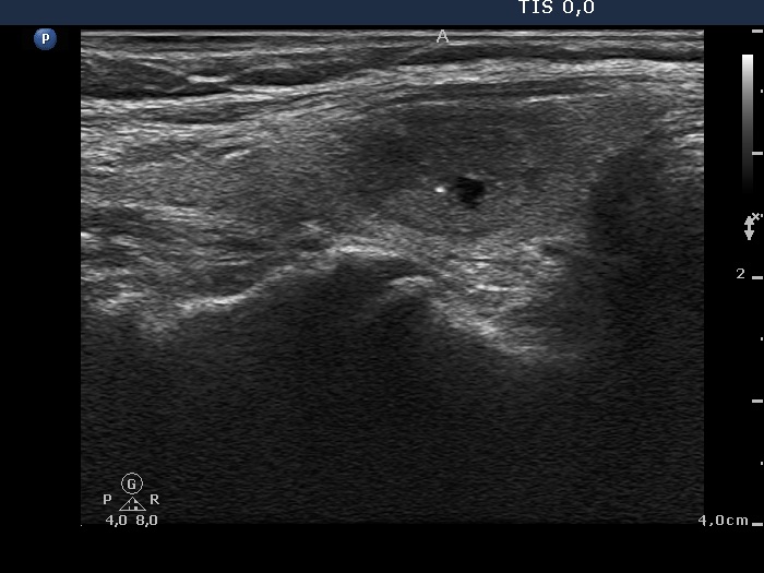
The synchronous presence of hyperechogenic lines and granules proves that these figures represent connective tissue while one in the ventral part does represent a colloid crystal.
|
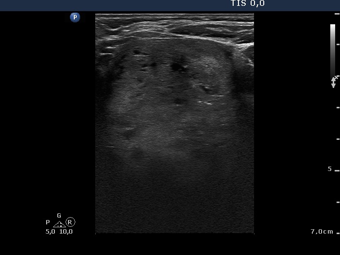
There are a few hyperechogenic granules within the small lesion and we can see two much thinner lines, as well. These figures might be either punctate echogenic foci or non-specific granules of a normal connective tissue.
|
| |
|
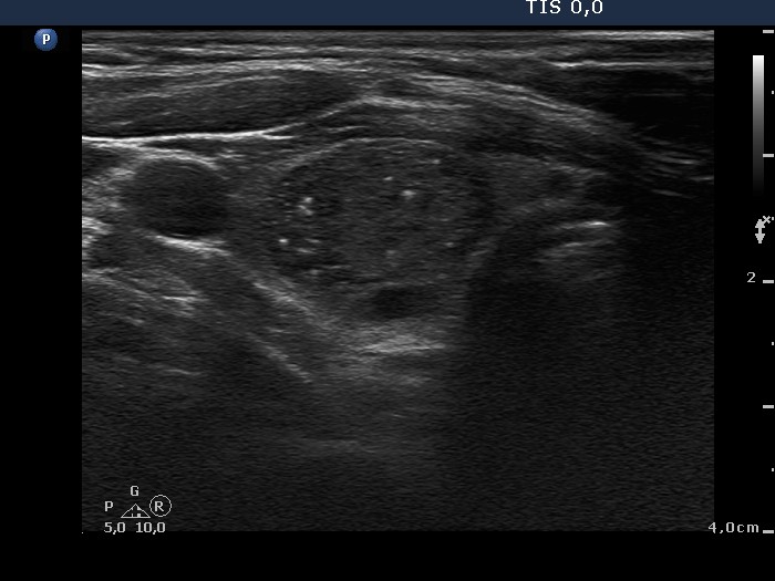
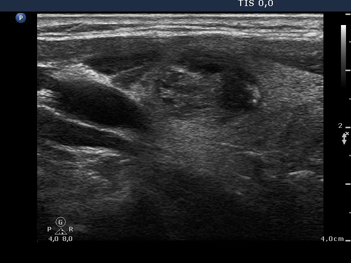
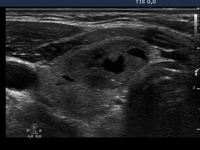
Follicular adenoma (histological diagnosis) - case cons 024 |
Benign hyperplastic nodules (histological diagnosis) - case 1091 |
 |
 |
 |
  |
Beside the very tiny granules and lines we find larger hyperechogenic granules, while the presence of similarly bright lines is spared. It is therefore ambiguous whether these figures are punctate echogenic foci or presentation of a connective tissue.
|
Several bright granules have dorsal pale tail therefore these and probably also the other granules lacking a tail belong to a comet-tail artifact subgroup.
|
| |
|
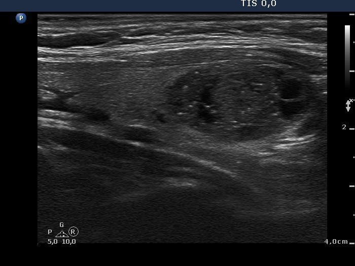
Hashimoto's thyroiditis with several cystic areas but without any nodules (histological diagnosis) - case 1365
|
 |
 |
 |
 |
There is no doubt of the origin of the hyperechogenic figure in the cystic lesion, this is a typical comet-tail artifact.
|
At first sight the interpretation of these figures seems to be difficult. However, this lesion is in the same patient whose other lesion is presented in the left images. It is reasonable to conclude that similarly bright figures within lesions presenting an identical echo pattern have the same origin.
|
| |
|
Hyalinizing trabecular adenoma (histological diagnosis) - case 510 |
Follicular carcinoma (histological diagnosis) |
 |
 |
 |
 |
Hyperechogenic granules coexist with lines - these are presentations partly of connective tissue partly comet-tail artifacts. (The analysis of video is clearly superior to the images in this case.)
|
The less bright granules and lines are presentations of connective tissue, while the four to five (upper image) and two or three (lower longitudinal scan) brighter tiny granules might correspond to punctate echogenic foci.
|
| |
|
Benign hyperplastic nodule (histological diagnosis) - case 1091 |
Papillary carcinoma (histological diagnosis) - case 1731 |
|
 |
 |
 |
Small granules dominate the nodule. At least two of them have dorsal narrowing tale. The remaining figures are similarly bright therefore these are very likely also comet-tail artifacts.
|
It is very difficult to categorize these figures. Considering the final diagnosis even the small pale figures might be punctate echogenic foci because there are no similarly pale lines. The two large bright granules are more typical forms of punctate echogenic foci. Note that there is a less hyperechogenic shadow not only dorsal but even ventral to the larger one. It means that this figure is not a comet-tail artifact but the granule is located in a minimally hypoechogenic parenchyma.
|
| |
|
Benign hyperplastic nodule (histological diagnosis) - case 1505 |
Benign hyperplastic nodule (histological diagnosis) - case 1449 |
 |
 |
 |
 |
Acoustic shadowing in the upper, horizontal view proves the presence of coarse calcification but most of the hyperechogenic granules are punctate echogenic foci.
|
Although several granules keep going dorsal and therefore comet-tail artifact has to be considered, at least part of these complex structures are composed of distinct granules which raises the possibility that these are in fact punctate echogenic foci.
|
| |
A multinodular goiter including a suspicious nodule (cytological diagnosis) - case cons 011
|
Benign colloid goiter (cytological diagnosis)
|
Suspicion of papillary carcinoma (cytological diagnosis) |
 |
 |
 |
 |
The coexistence of lines and granules of similar brightness in the most hypoechogenic lesion (left images) proves that these figures are connective tissue. The less hypoechogenic lesion in the right upper image also has non-specific figures.
The least hypoechogenic lesion (left images) presents only granules which correspond to punctate echogenic foci.
|
| |
|
Follicular adenoma (histological diagnosis) - case 1056 |
Papillary carcinoma (histological diagnosis) - case 1074 |
 |
 |
 |
 |
 |
 |
The presentation of the granules is similar in these cases. However the benign lesion has several granules with dorsal tail, therefore these correspond more likely to comet-tail artifacts than to punctate echogenic foci. On the other hand, the complex structures in the malignant nodules are composed of multiple granules. It is worth comparing the figures marked with arrows.
|
| |
|
Benign hyperplastic nodule (histological diagnosis) - case 837 |
Papillary carcinoma in Hashimoto's thyroiditis (histological diagnosis) - case 455 |
 |
 |
 |
 |
The bright granules partly have dorsal tail, these are therefore comet-tail artifacts. Moreover, this is a small cyst; therefore the granules within the fluid cannot be anything other than colloid crystals.
|
There are large bright granules in the lower, longitudinal image. With this older equipment these figures might be punctate echogenic foci.
|
| |
|
Follicular adenoma (histological diagnosis) - case 932 |
Benign hyperplastic nodules (histological diagnosis) - case 907 |
 |
 |
 |
 |
The coexistence of lines and granules of similar brightness proves that these figures are the connective tissue.
|
Hyperechogenic lines of various levels of brightness dominate the nodule. These figures are presentations of the connective tissue.
|
| |
|
Benign hyperplastic nodule (histological diagnosis) - case 444 |
Oxyphilic adenoma (histological diagnosis) - case 368 |
 |
 |
 |
 |
Beside non-specific figures (pale tiny granules and lines) corresponding to connective tissue the lesion has two larger granules. These might be punctate echogenic foci, however on video it is evident that almost similarly bright lines are present; therefore the large granules likely correspond also to a connective tissue.
|
There are three granules in the upper one in the lower image. They are punctate echogenic foci.
|
| |
|
Benign hyperplastic nodule (histological diagnosis) - case 1209 |
Follicular adenoma (histological diagnosis) - case 1206 |
 |
 |
 |
 |
There are different types of hyperechogenic figures including colloid crystals without the typical tail in the cystic area, figures caused by a posterior back wall enhancement and a connective tissue in the presence of bright granules and lines in the solid part of the nodule.
|
The two bright spots must be held as punctate echogenic foci.
|
Benign cystic-colloid goiter (cytological diagnosis) - case 1490 |
Hashimoto's thyroiditis (cytological diagnosis) - case 1500 |
 |
 |
 |
 |
Several hyperechogenic granules present a dorsal gradually paling tail; therefore these are comet-tail artifacts.
|
Except for one or two vague lines there are only hyperechogenic granules within the lesion. They lack dorsal tail therefore they cannot be categorized other than punctate echogenic foci.
|
| |
|
Benign colloid goiter (cytological diagnosis) - case cons 029 |
Papillary carcinoma (histological diagnosis) - case 1777 |
 |
 |
 |
 |
The solid part of the nodule has both hyperechogenic granules and lines, i.e. connective tissue. There are typical colloid crystals in the cystic area.
|
The lesion presents two or three relatively bright granules. In the absence of similarly bright lines these are punctate echogenic foci.
|
| |
|
Follicular adenoma (histological diagnosis) - case 1406 |
Medullary carcinoma (histological diagnosis) |
 |
 |
 |
 |
There are several typical forms of comet-tail artifacts in the upper image. There are two hyperechogenic figures, and the larger one is very difficult to categorize. Considering the presence of comet-tail artifacts these ambiguous figures probably belong also to the same subgroup of figures.
|
There are several bright tiny hyperechogenic granules which have to be grouped as punctate echogenic foci. The large, complex figure in the central part of the lower image corresponds to amyloid deposit: there are small bright hyperechogenic granules within a less hyperechogenic, irregularly shaped structure.
|
| |
|
Benign hyperplastic nodule (histological diagnosis) - case 186 |
A cyst has been treated with ethanol for 6 years - case 187 |
 |
 |
 |
 |
The lesion has non-specific lines and granules. There are two more bright granules, one in the upper-ventral and another in the lower part of the lobe. These might be punctate echogenic foci.
|
Bright granules coexist with lines - the typical presentation of proliferation of connective tissue. (The lines are better seen in the video.)
|
| |
|
Benign follicular proliferation - case 11 |
Papillary carcinoma - case 979 |
 |
 |
 |
 |
Hyperechogenic lines and granules coexist, these figures represent connective tissue. In the presence of the acoustic shadow the lesion has to contain coarse calcifications, too.
|
The tumor has an interrupted peripheral calcification and a few punctate echogenic foci, as well. There is a hyperechogenic large complex structure in the left; the horizontal view is difficult to judge. It might be ragged tissue containing punctate echogenic foci.
|
| |
|
Metastasis of a kidney carcinoma to the thyroid - case 1574 |
Benign hyperplastic nodule (histological diagnosis) - case 1582 |
 |
 |
 |
 |
Both images show one relatively large granule which correspond to punctate echogenic foci.
|
Parts of the hyperechogenic granules are located in cystic area, while others are found in the solid part of the lesion. The latter might be punctate echogenic foci.
|