
|
|
100 consecutive patients with thyroid nodule - Case 11.
|
|


Clinical presentation: hypertension and diabetes mellitus were diagnosed in a 62-year-old woman. During her evaluation a nodular goiter was discovered.
Palpation: the lower pole of the right thyroid was firm on palpation.
Functional state: euthyroidism (TSH 1.03 mIU/L).
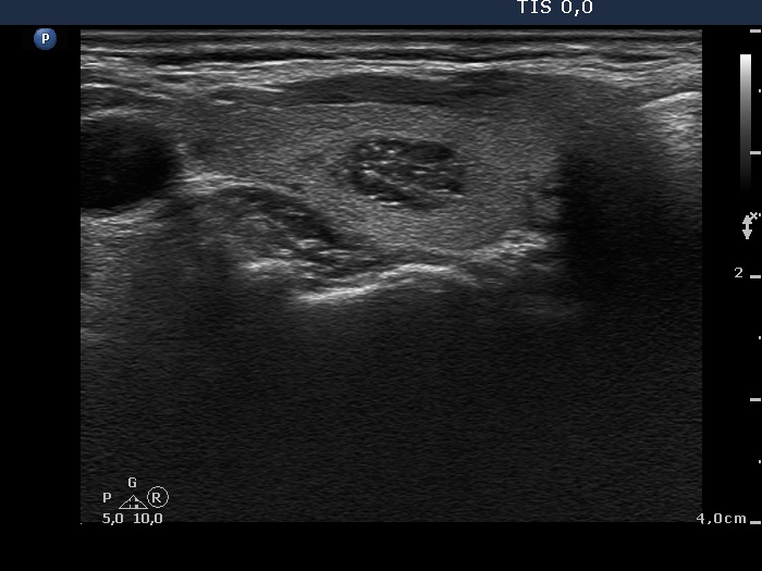
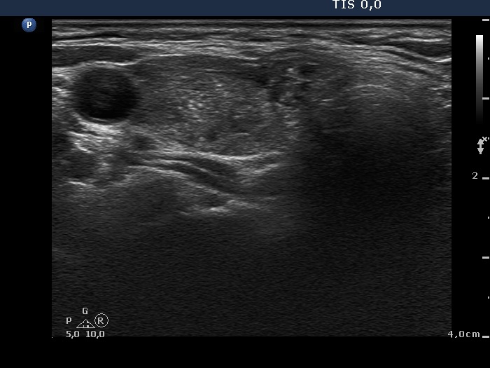
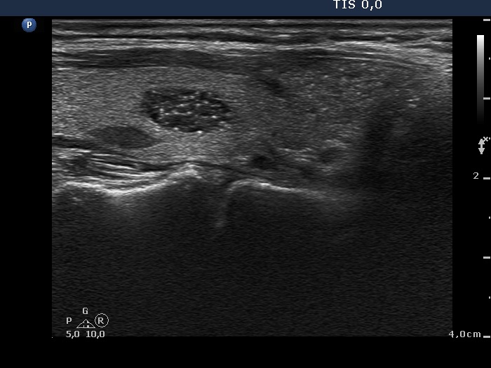
Ultrasonography: the thyroids were echonormal. There was a hypoechogenic nodule in the central part of the right lobe and a larger moderately hypoechogenic lesion presenting ill-defined borders and microcalcifications in the lower pole of the right thyroid. A third nodule was found in the left lobe.
The nodules in the right lobe were aspirated. Cytology of the nodule in the middle part of the lobe was benign. The cytology of the lesion in the lower pole of the right lobe corresponded to follicular proliferation.
Considering the echo pattern we gave a common ultrasound-cytological diagnosis: benign colloid goiter and suspicion of a thyroid neoplasia with around 20% risk of malignancy, nodule in the middle and in the lower third of the right lobe, respectively.
Histopathological result is not known, yet.
Comment. The cytological pattern itself is not very suspicious. Although the ratio of isolated microfollicles to those located in larger sheets is a bit higher, other cytological signs of a follicular tumor are lacking. On the microscopic analysis, the risk of malignancy cannot be greater than 1-2 %.
However, if we take the sonographic pattern into account, the possibility of a carcinoma, i.e. a follicular variant of papillary carcinoma has to be considered.
As regards the ultrasound presentation there were two features which increased the risk of malignancy: the presence of microcalcifications and the ill-defined borders of the lesion.