
|
|
100 consecutive patients with thyroid nodule - Case 12.
|
|


Clinical data: a 48-year-old man requested evaluation of a lump in the right thyroid which was discovered for 3 weeks.
Palpation: an elastic nodule in the right lobe.
Functional state: euthyroidism (TSH 2.17 mIU/L).
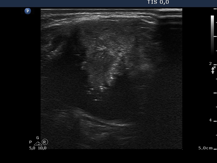
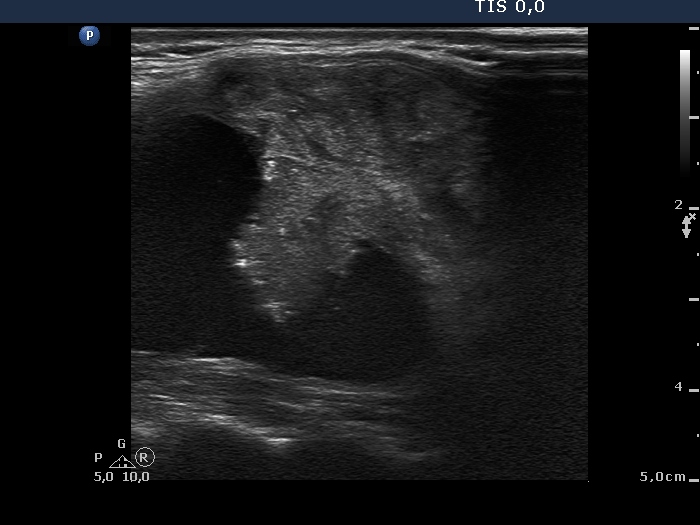
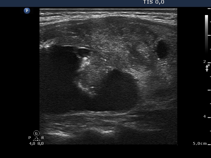
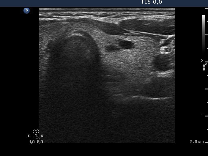
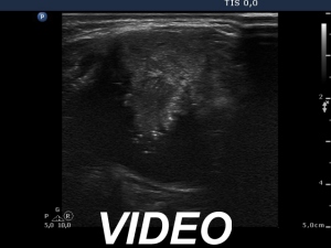
Ultrasonography: the thyroid was echonormal. There was a mixed, peripheral-type cystic nodule in the right lobe while a smaller cystic lesion was found in the left thyroid. The former presented signs of perinodular, type 2 vascular pattern and an echonormal solid part.
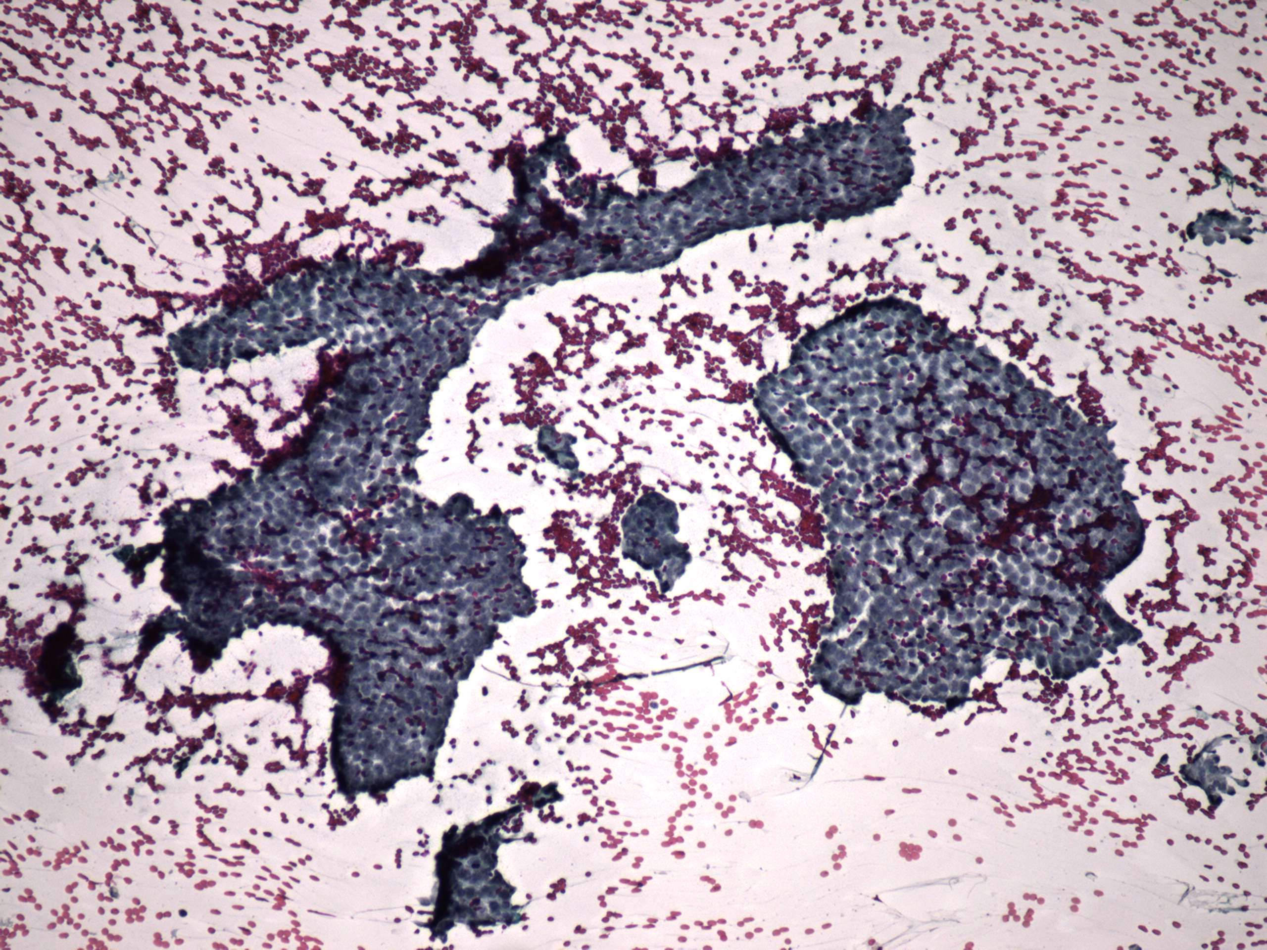
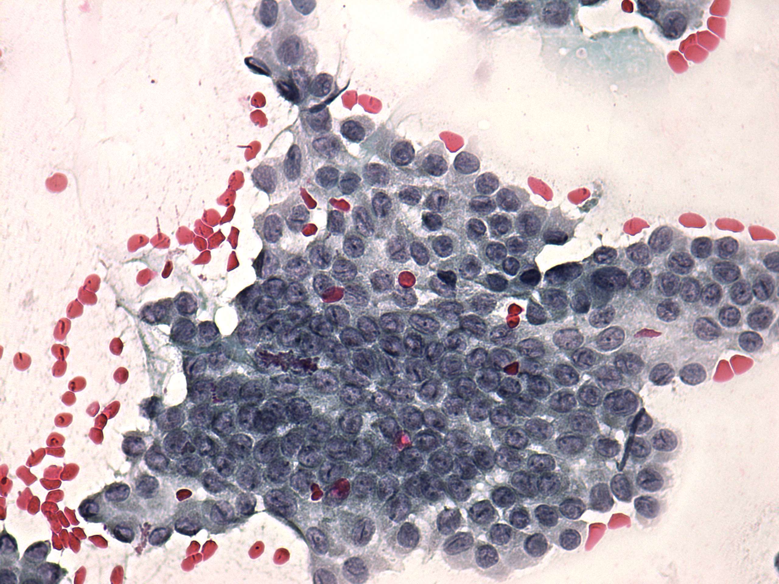
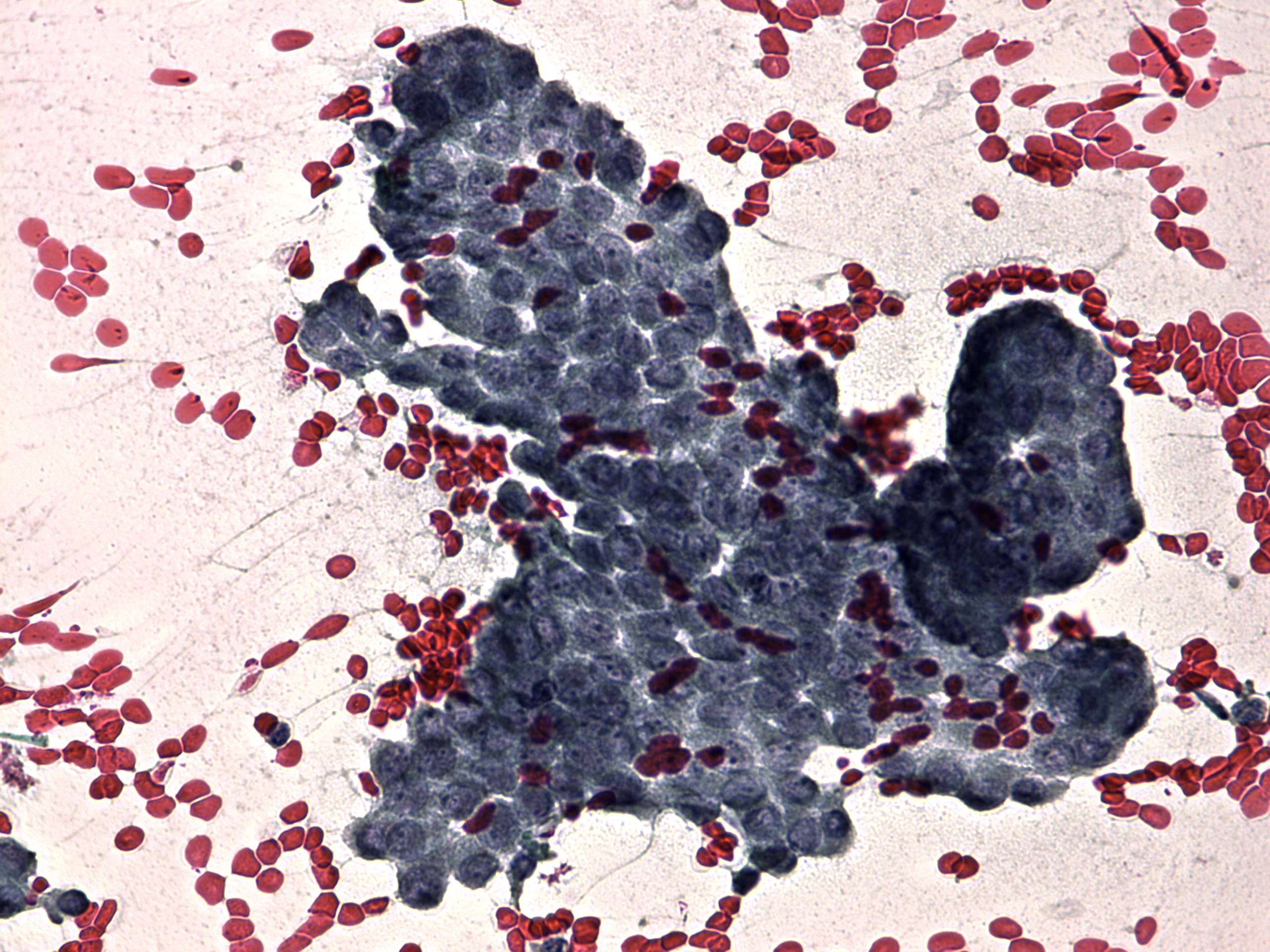
On FNAC 0.5 ml bloody fuid was aspirated. There were only macropahges and a few isolated follicular cells on the smear.
Combined ultrasound-cytological diagnosis: thyroid cyst with not greater than 1% risk of malignancy.
Comments:
-
As regards the ultrasound presentation there was one feature which increased the risk of malignancy: the peripheral-type of the cyst. On the other hand, other features decreased the likelihood of carcinoma: the cyst presented blunt angle and the vascularization was not increased, the solid part was almost echonormal and did not contained microcalcification.
-
Although the cytology itself was not diagnostic, considering the ultrasound presentation we gave the above diagnosis.