
|
|
Parathyroid lesions - Case 9: an intrathyroidal parathyroid adenoma
|
|

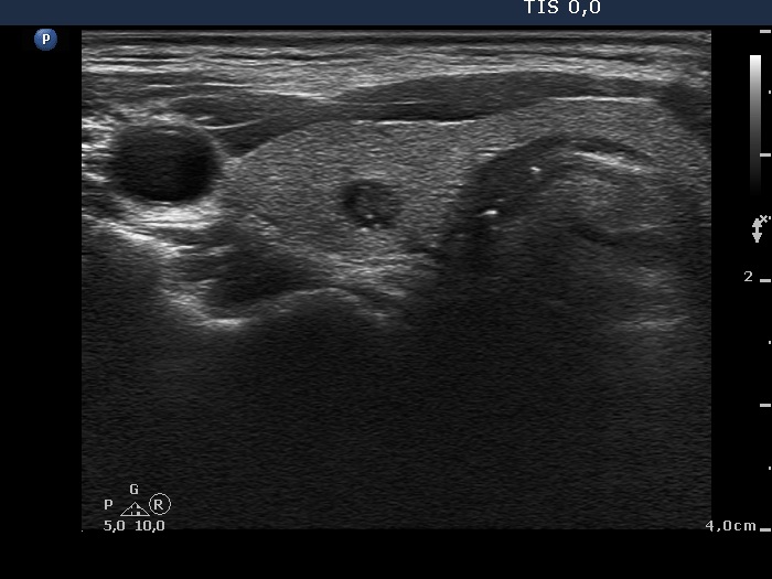


Clinical presentation: A 57-year-old woman was referred for evaluation of a hyperparathyroidism and elevated serum calcium level.
Palpation: no abnormality.
Laboratory tests: TSH 3.70 mIU/L, calcium 3.08 mM/L, phosphorus 0.67 mM/l, parathormone 177.4 pg/mL (normal value: 15-66).
Ultrasonography: The thyroid was echonormal. There was a small hypoechogenic lesion in the upper central part of the right lobe. The lesion presented hyperechogenic granules. We could not find any discrete abnormalities outside the thyroid.
Aspiration cytology of the lesion resulted in benign lesion which might correspond either to a parathyroid adenoma or a benign thyroid lesion.
Wash-out thyroglobulin was low (9.08 mg/L), while wash-out parathormone resulted in 203 pg/mL. Serum thyroglobulin was 9.84 mg/L.
Our final diagnosis was parathyroid adenoma.
Scintigraphy was equivocal as regards the lesion within the thyroid, while there were no other positive masses on MIBI scintigram.
Right lobectomy was performed. Histopathology: benign parathyroid adenoma within an intact thyroid.
Comments.
-
The least frequent location of a parathyroid is the intrathyroidal one. The only chance to recognize an intrathyroidal lesion as a parathyroid if we are aware of hyperparathyroidism.
Moreover, even considering the hyperparathyroidism, the likelihood that this intrathyroidal lesion is of parathyroid origin was significantly lower than the chance of a thyroidal origin. -
The wash-out technique was very useful.