
|
|
Parathyroid lesions - Case 10: A parathyroid adenoma with a deceptive ultrasound and cytological presentation
|
|


Clinical data: a 40-year-old woman was referred for evaluation of a nodule detected by the patient herself. Aspiration cytology resulted in AUS-FLUS (Bethesda) in another institute.
Palpation: a not firm lesion in the lower-lateral part of the right thyroid bed.
Laboratory test: TSH 2.40 mIU/L.
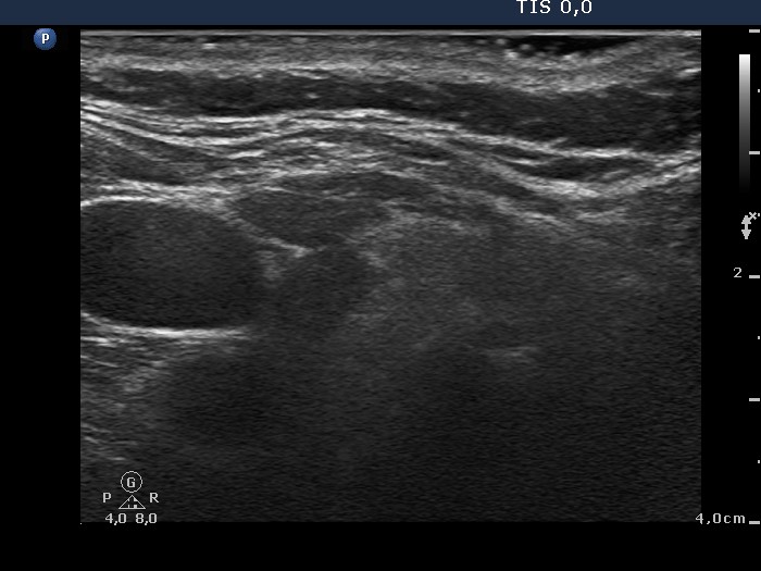
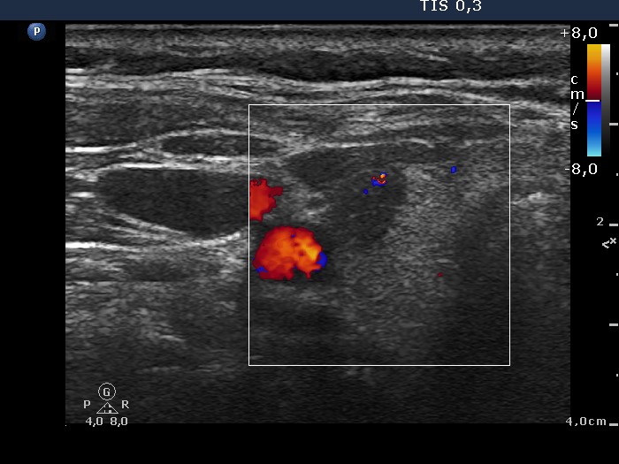
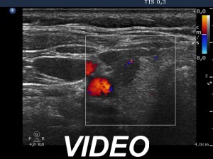
Ultrasonography: the thyroids were echonormal, the left lobe was inhomogeneous. There was a hypoechogenic lesion in the upper ventrolateral part of the right lobe. The vascularization was not specific.
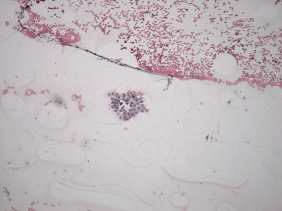
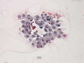
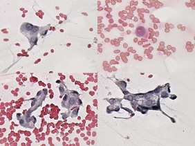
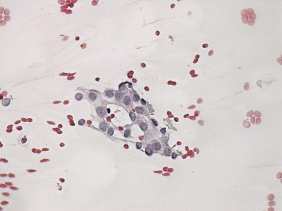
Cytology. There were only few cell groups on the smear. The cells had abundant cytoplasm and presented vacuolization. Several nuclei in the largest cluster displayed inclusion. Our diagnosis was suspicion of papillary carcinoma.
A right lobectomy was performed. Histopathology disclosed parathyroid adenoma.
Comments.
-
The location of the enlarged parathyroid was unusual it seems to be located within the thyroid.
-
An even greater concern arises viewing the cytological presentation. Although there were only a few cell groups on the smear, the frequency of inclusion was deceptively high.
-
It is very hard to reconsider our cytological diagnosis. Moreover, the ultrasound presentation was of help not to give a definitive diagnosis of papillary carcinoma because the lesion did not presented any signs of malignancy.