
|
|
Benign nodular hyperplasia - Case 16.
|
|


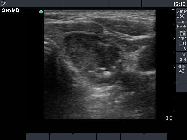


First investigation (first row of images)
Clinical presentation. A 62-year-old man detected a nodule in her thyroid 2 months earlier.
Functional state: euthyroidism (TSH 1.26 mIU/L).
Ultrasonography. There was a mixed cystic-minimally hypoechogenic nodule in the left lobe.
Aspiration cytology: 3 mL brown cystic fluid was aspirated, thereafter the solid part of the nodule was aspirated. There were only macrophages and red blood cells on the smear.
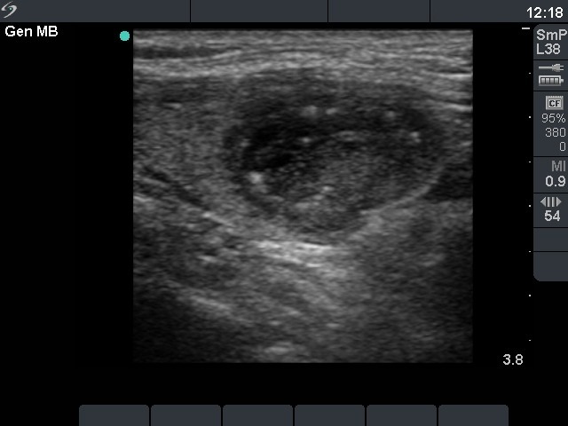
Second investigation (second and third rows of images)
Clinical presentation. The patient told us that the cyst has refilled several weeks after the aspiration.
Combined sonographic-cytological diagnosis: "the risk of papillary cancer is in the range of 5-15%. (Atypia of unknown significance.)"
Histopathology disclosed benign hyperplastic nodular goiter.
Comments.
-
On our opinion, this case is a typical example of atypia of unknown significance (category AUS of Bethesda system). The atypia may be explained either by degenerative changes or by a papillary carcinoma.
-
The clinical and sonographic properties, i.e. the recurrence of the cyst, the presence of microcalcifications increased the risk of papillary cancer.
-
This case was part of our prospective study testing the usefulness of category AUS of Bethesda system. Considering the risk of malignancy, the patient decided to undergo surgery instead of follow-up examinations.