Benign nodular hyperplasia - Case 16.
Second examination one year later (ultrasonographic picture 2)

|
|
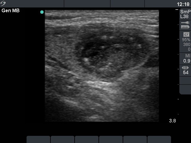
Left lobe, longitudinal scan. This is a moderately hypoechogenic, inhomogeneous nodule.

|
|
|
Left lobe, longitudinal scan. This is a moderately hypoechogenic, inhomogeneous nodule.