Benign nodular hyperplasia - Case 16.
Second examination one year later (ultrasonographic picture 1)

|
|
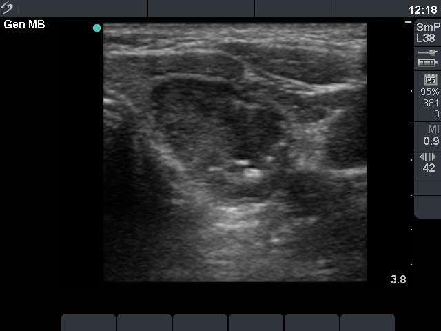
Left lobe. horizontal scan. A moderately hypoechogenic nodule with smaller and larger hyperechogenic foci.

|
|
|
Left lobe. horizontal scan. A moderately hypoechogenic nodule with smaller and larger hyperechogenic foci.