
|
|
Follicular adenoma - Case 11.
|
|


Clinical data: A 32-year-old man was referred for an evaluation of a thyroid nodule which was discovered on ultrasonographic examination indicated because the patient felt swelling in the neck for several months.
Palpation: a firm nodule in the right lobe.
Functional state: euthyroidism with TSH 3.11 mIU/L, FT4 13.9 pM/L.
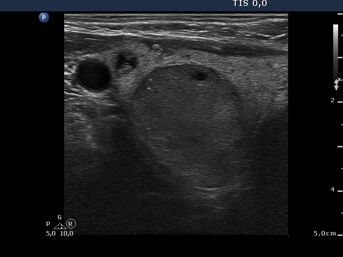
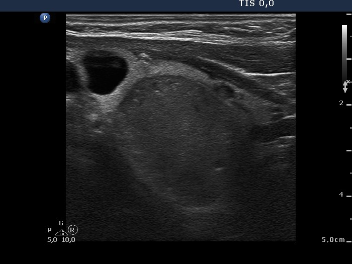
Ultrasonography: The thyroid was echonormal and contained several small, moderately hypoechogenic and hypoechogenic lesions. One of them in the left lobe contained hyperechogenic granules. There was a large hypoechogenic nodule in the right lobe displaying halo, microcalcifications and signs of perinodular blood flow.
Cytology was performed from the nodule is the right lobe (first row of cytological images) and from the lesion presenting microcalcifications in the left lobe (second row of cytological images).
Cytological diagnosis form the right nodule: follicular tumor with greater than the average risk of malignancy.
Cytological diagnosis from the left lesion: with great probability a benign lesion with signs of hormonal influences.
We performed scintigraphy which demonstrated a cold nodule in the right lobe. Anti-TPO and TSAb tests were negative.
Histopathological diagnosis: follicular adenoma with significant cellular atypia according to the large nodule in the right thyroid. Benign hyperplastic nodules in both lobes.