
|
|
Benign nodular hyperplasia - Case 4.
|
|


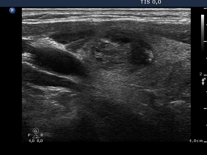

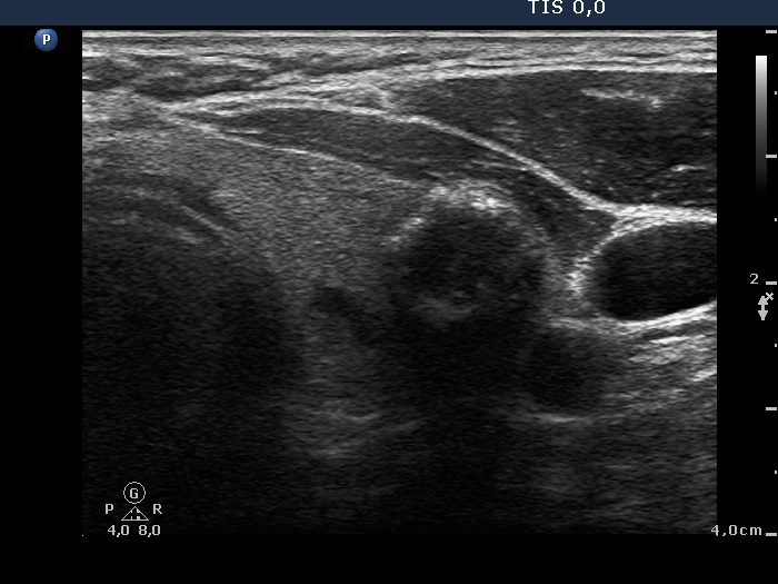


Clinical presentation: A 49-year-old man was referred for an evaluation of a nodular goiter detected on screening.
Palpation: The left lobe was enlarged and multiple nodules were palpable.
Functional state: euthyroidism with TSH-level 0.65 mIU/L.
Ultrasonography: The right lobe had a moderately hypoechogenic, the left had multiple nodules.
Cytology was performed from the nodule in the right lobe, from the calcified part and from the moderately hypoechogenic nodule in the left lobe. The cytological patterns were identical in all three cases and yielded benign, colloid goiter.
The patient was operated because of the size of the left lobe.
Histopathology disclosed benign, hyperplastic nodules.