Benign nodular hyperplasia - Case 4. (ultrasonographic picture 4)
|
|

|
|
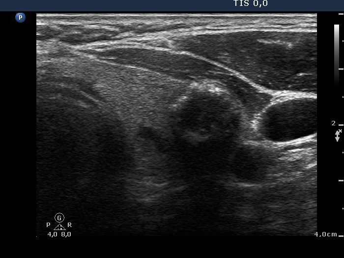
Left lobe, horizontal view. A nodule with coarse calcification is demonstrated in the image.

|
|
|
|
Left lobe, horizontal view. A nodule with coarse calcification is demonstrated in the image.