
|
|
Benign nodular hyperplasia - Case 5.
|
|


Clinical presentation: a 40-year-old woman was referred for an evaluation of a nodular goiter known for years. She had neck discomfort and occasional difficulties in swallowing.
Palpation: the right lobe was enlarged and a nodule was palpable.
Functional state: euthyroidism with TSH-level 1.08 mIU/L.
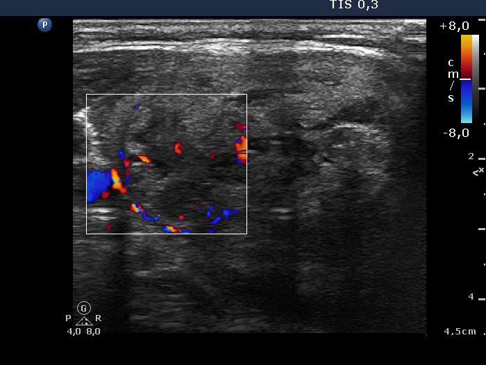
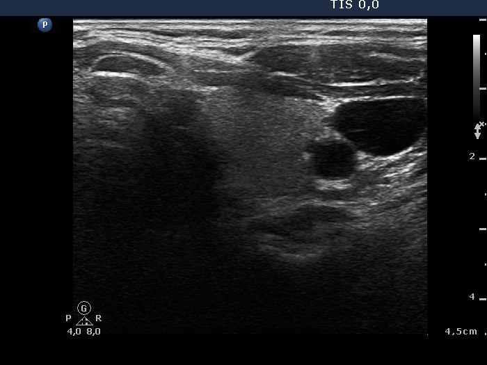
Ultrasonography: the right thyroid contained a mixed nodule with hypoechogenic, echonormal and cystic components. The nodule displayed halo sign and both perinodular and intranodular blood flow.
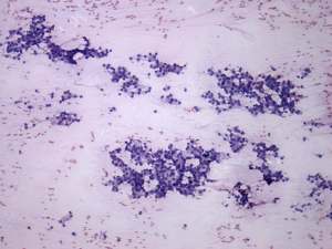
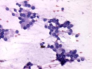
Cytology: there was no colloid in the background. A great proportion of follicular cells were found in normo- and microfollicles. The nuclei were alike in size and presented prominent nucleoli. A few cells contained inclusion while a relatively large proportion of nuclei contained groove.
Cytological diagnosis: suspicion of follicular variant of papillary cancer.
Histopathology: disclosed benign, hyperplastic nodules in the right lobe.
Comment: this case is a typical example of atypia of unknown significance. Although this lesion proved to be hyperplastic nodular goiter, this sonographic pattern, i.e. an intact thyroid in one side while a large nodule with halo sign in the other side is a specific pattern of a follicular tumor.