
Benign nodular hyperplasia - Case 5. (ultrasonographic picture 3)
|
|

|
|
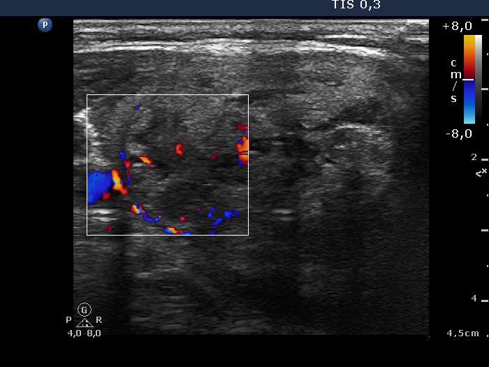
Right lobe, horizontal view, color Doppler mode. Combined type 2 and type 3 vascular pattern, ie. the nodule displays both perinodular and intranodular blood flow.