
|
|
Lymph nodes in the neck - Case 3.Benign, bacterially infected lymph node
|
|


Clinical data: A 16-year-old girl was referred for evaluation of a thyroid nodule discovered on evaluation of an upper airway infection. Her complaints began three months ago and lasted for 2 weeks. 5 day before present investigation she ran a temperature. A bacterial infection was diagnosed with elevated CRP levels. Antibiotics was started two days before our investigation. The fever has stopped.
Palpation: There was a tender mass in the right submandibular area. There was no abnormality in the thyroid.
Functional state: euthyroidism (TSH 3.13).
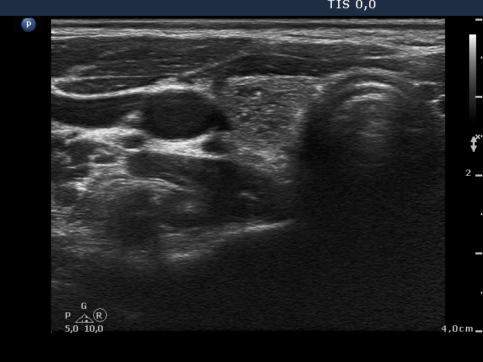
Ultrasonography: The thyroid was echonormal. There was a moderately hypoechogenic inhomogeneous nodule in the lower pole of the right lobe. There were two lymph nodes next to each other above the right thyroid in the submandibular area. The lymph nodes presented a regular hilum.
Cytology was performed from the nodule in the right thyroid and resulted in a benign colloid goiter.
Follow-up investigation one year later. The size of the thyroid nodule remained unchanged while the lymph nodes have disappeared.
Comments.
-
This is the typical presentation of a bacterially infected lymph node. In most of these cases we find multiple lymph nodes next or very close to each other. The presence of ill-defined hypoechogenic areas within the lymph nodes is an even more important sign of a bacterial lymphadenitis.
-
It is edifying to analyze the hyperechogenic figures in the thyroid lesion. While examining the patient, I interpreted these as microcalcifications. However, by preparing the case study I had to regroup these figures. These are in fact presentations of connective tissue and colloid crystals. (See the footnotes of the relevant images.)