
Lymph nodes in the neck - Case 3. (ultrasonographic picture 2)
Benign, bacterially infected lymph node
|
|

|
|
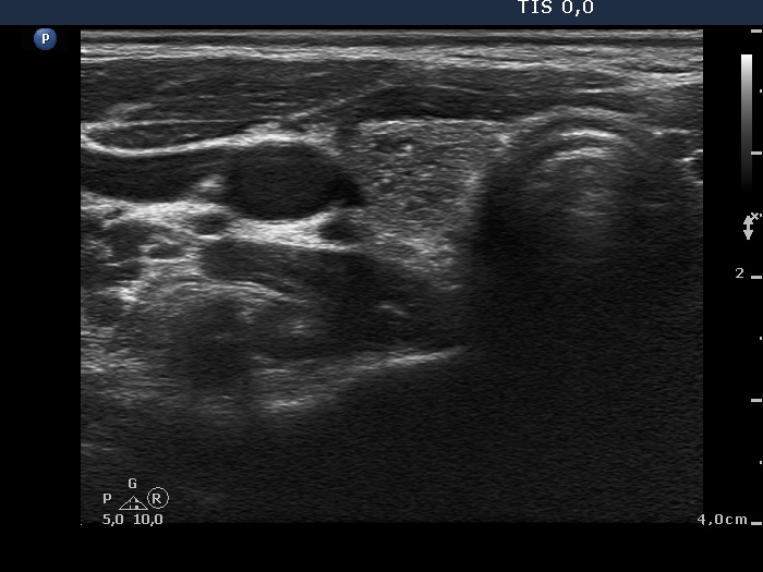
Lower pole of the right lobe, horizontal scan. There is a moderately hypoechogenic nodule presenting hyperechogenic granules. Considering the coexistence of hyperechogenic lines, these figures are more likely signs of proliferation of connective tissue or back wall enhancement.