
|
|
Benign nodular hyperplasia - Case 49.
|
|


Clinical presentation: A 78-year-old woman was referred for evaluation of a goiter known for decades. The thyroid slowly increased in size and the patient already suffered from dyspnea and dysphagia in the last 6 months. Previous scintigraphy indicated a cold nodule in the left lobe.
Palpation: a very enlarged, firm nodule with uneven surface in the left lobe.
Functional state: euthyroidism (TSH 0.53 mIU/L).
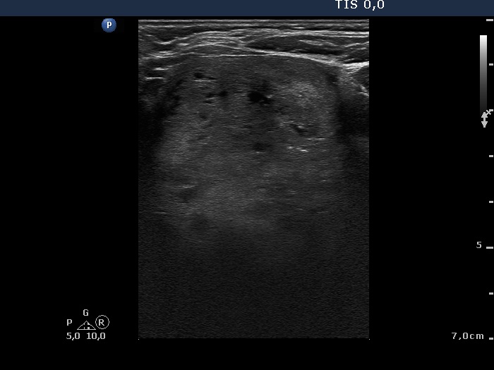
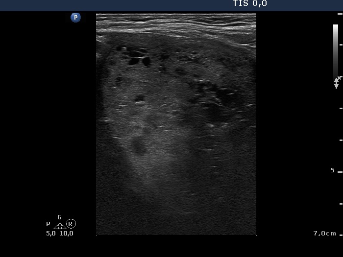
Ultrasonography: The right lobe was normal in size and contained several small nodules with different echogenicities. The left lobe was consisted of a large nodule composed of smaller lesions. It contained numerous hyperechogenic figures which corresponded to back wall figures and presentation of connective tissue. The left lobe spread retrotracheal and substernally. The lower pole of the lobe was not clearly visible while the patient was swallowing.
Cytological diagnosis: benign follicular proliferation with less than 1% of an estimated risk of carcinoma.
Neck and upper mediastinal CT scan was performed which disclosed that the left thyroid was spread 12 cm below the level of the clavicle.
Left lobectomy was performed. Histopathology disclosed benign hyperplastic nodules and a solitary focus of papillary carcinoma with 2 mm in maximal diameter.
Comments.
-
It is worth analyzing the hyperechogenic granules in video. Nomen est omen, they have a tail which is less well reproducible in an image than in video.
-
The cytological pattern was identical to a follicular tumor. Nevertheless, we gained the smear from a multinodular goiter which presented neither halo sign nor perinodular vascularity. Therefore, a combined cytological-sonographic diagnosis was benign follicular proliferation instead of a follicular tumor. The correct preoperative diagnosis was of great importance. If the diagnosis would be follicular tumor than the patient would undergo on total thyroidectomy because her cardiac status was wrong and a repeat surgery had to be avoided if final histopathology would disclose carcinoma.