
Benign nodular hyperplasia - Case 49. (ultrasonographic picture 4)
|
|

|
|
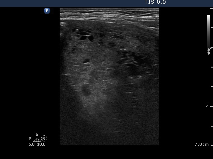
Left lobe, longitudinal scan. The nodule presents numerous hyperechogenic granules. These figures are mostly elongated and partly related to ventral cystic areas. It means that these are back wall figures and/or presentations of connective tissue.