
The operated thyroid - Case 23. A patient after a lobectomy
One year after surgery (ultrasonographic picture 8)
|
|

|
|
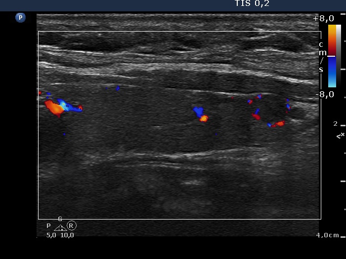
Left lobe, longitudinal scan, color Doppler mode. The small lesion presents a type 2 vascular pattern.