
Differentiation of cystic thyroid lesions - case 126
First session of sclerotherapy (ultrasonographic picture 2)
doi: 10.24390/thyrocase126.1.02

|

|
 |
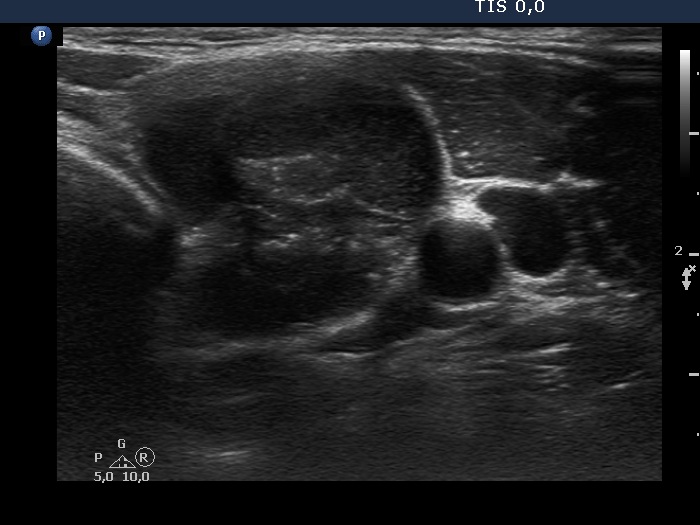
Lower part of the left lobe, horizontal scan. The lower pole of the nodule has a solid part with numerous echogenic figures. These include figures caused by posterior cystic enhancement, non-specific granulations. In a few granules punctate echogenic foci (microcalcifications) has to be considered.


