
Differentiation of cystic thyroid lesions - case 126
First session of sclerotherapy (ultrasonographic picture 6)
doi: 10.24390/thyrocase126.1.06

|

|
 |
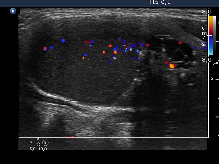
Left lobe, longitudinal scan, color Doppler mode. The pattern of the upper 4/5 of the lesion is deceptive: this part of the nodule contained cystic fluid, so the blue and red spots do not correspond to vessels.


