
Graves' disease - Case 1. (ultrasonographic picture 4a)
|
|

|
|
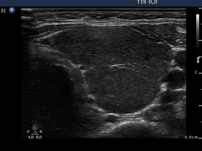
Left lobe, horizontal scan. This image demonstrated the substructure of a thyroid lobe. We can identify three pseudolobules separated by fibrotic tissue.