
Graves' disease - Case 1. (ultrasonographic picture 4b)
|
|

|
|
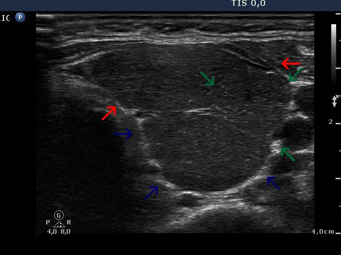
Left lobe, horizontal scan. This image demonstrated the substructure of a thyroid lobe. The three pseiudolobules are marked with different arrows.