
|
|
Differentiation of cystic thyroid lesions - Case 1295doi: 10.24390/thyrocase1295.00
|

|


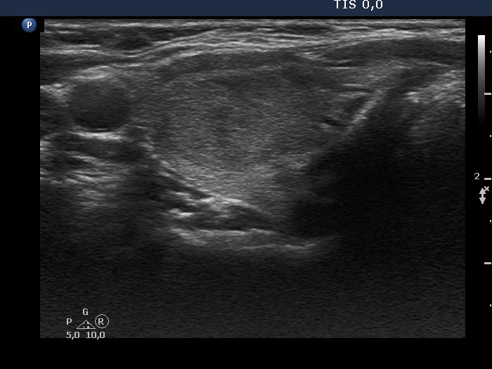
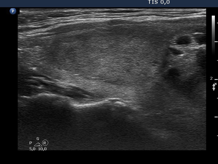
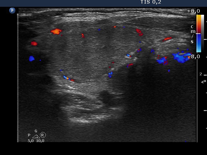
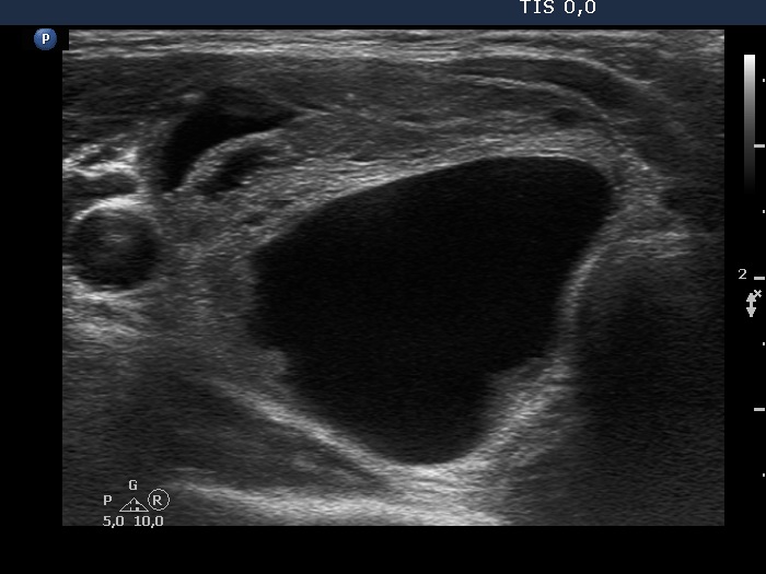
Clinical presentation: A 51-year-old woman had had a myocardial infarction for a year. On evaluation a subclinicial hyperthyroidism and thereafter an autonomously functioning adenoma and a cystic nodule were diagnosed. She was sento to aspiration cytology before surgery.
Palpation: The right lobe was enlarged and nodular on palpation.
Result of blood tests: TSH 1.94 mIU/L, FT4 10.8 pM/L on daily 5 mg methimazole.
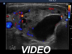
Ultrasonography. The thyroid was echonormal. There was minimally hypoechogenic nodules in the upper part of the left lobe. The lesion has perinodular blood flow. A larger cystic nodule was found in the lower part of the right lobe. Thorough analyis revealed that this was a central-type cystic lesion with one large and sveral smaller cystic chambers.
Besides macrophages there were only a few isolated follicular cells on the smear. A combined ultrasound-cytological diagnosis was given: cystic lesion with less than 1% risk of malignancy.
Right lobectomy was performed. Histopathology disclosed follicular adenoma in both nodules.


