
|
|
Oxyphilic adenoma - Case 21.
|
|


Clinical presentation: a 44-year-old woman requested a second opinion. She discovered a nodule for 2 years. The nodule slowly increased in size. On evaluation performed in another institute the final report was thyroid carcinoma with great probability and total thyroidectomy was planned.
Palpation: a firm nodule in the right thyroid bed.
Functional state: euthyroidism (TSH 3.15 mIU/L).
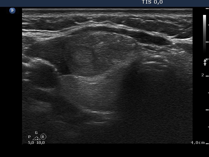
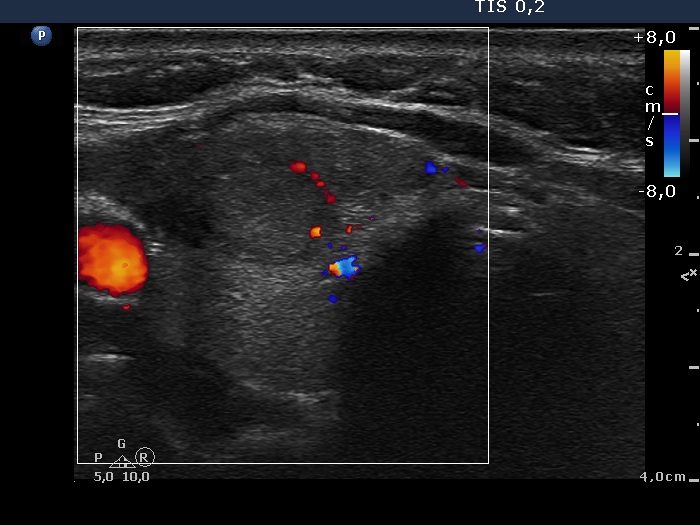
Ultrasound. The thyroid was echonormal. There was a moderately hypoechogenic mass located in the ventral surface of the right lobe. It was equivocal whether the lesion was within or outside the thyroid.
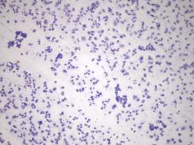
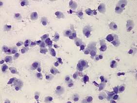
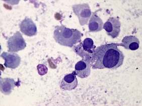
Cytology was performed from the nodule in the right lobe. The cellular smear showed round to polygonal cells with well-defined cell borders. Dissociated cells predominated the smear. The cells presented abundant cytoplasm with granulations.
Three possibilities had to be considered. An oxyphilic thyroid tumor, an oxyphilic parathyroid tumor and medullary carcinoma.
Wash-out thyroglobulin was above 478 microgram/L.
Our final diagnosis was Hürthle-cell tumor.
A right lobectomy was performed. Histopathology disclosed oxyphilic cell adenoma.
Comments.
-
Although the risk of a medullary carcinoma cannot be excluded on the cytological pattern, taking the ultrasound presentation into account, the risk of a medullary carcinoma was very low. Medullary carcinoma is almost always presented in the form of more hypoechogenic nodule.
-
The ultrasound appearance of the lesion was unusual. We could not decide whether this lesion is within or outside the thyroid. Although more than 80% of such cases proves to be of thyroidal origin, additional tests are required to decide te issue.
-
The result of wash-out decided the question.