
|
|
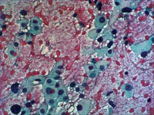
Oxyphilic adenoma - Case 19.
|
|


Clinical data: a 47-year-old woman was referred for an evaluation of a newly discovered nodule.
Palpation: a moderately enlarged right lobe. Multiple nodules were palpable in both lobes.
Functional state: euthyroidism (TSH-level 1.74 mIU/L).
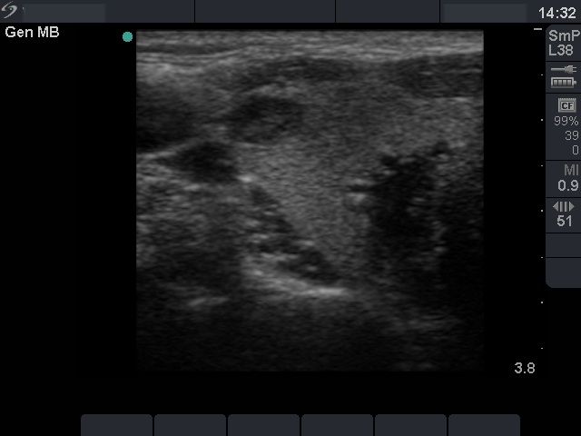
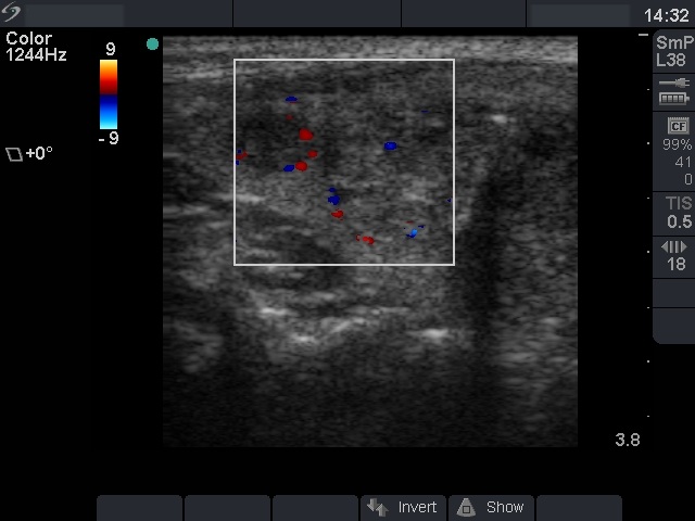
Ultrasonography: the basic echo structure of the thyroid was normal. There were three lesions in the right, and one lesion in the left lobe. The hypoechogenic one in the upper pole of the right lobe was aspirated.
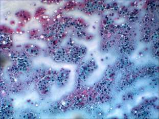
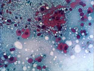
Cytological picture: no colloid in the background. Thyrocytes occur in loose clusters and dissociated. Almost all follicular cells exhibit oxyphilic metaplasia with prominent nucleoli. Many cells contain intranuclear grooves, some of them intranuclear inclusions. Thyrocytes exhibit pronounced pleomorphism.
Cytological diagnosis: oxyphilic variant of papillary cancer.
Histopathology: oxyphilic adenoma corresponding to the nodule in question. Hyperplastic nodules corresponding to the remaining ones.
Comment: this was one of our two cases where the FNAC was definitively false positive. The problem lies in the interpretation of nuclear grooves and inclusions in the case of oxyphilic cells. Any Hürthle-cell may contain inclusions and grooves - irrespectively from the origin of the lesion. This is the only situation where the specificity of these nuclear details is low. The pleomorphism or the nuclear crowding has only little relevance in the case of oxyphilic cells irrespectively of their inflammatory or tumorous origin.