
|
|
Oxyphilic adenoma - Case 18.
|
|


Clinical data: a 48-year-old woman was referred for an evaluation of a thyroid nodule that had been discovered a year earlier.
Palpation: a solitary nodule was palpable in the right lobe.
Functional state: euthyroidism (TSH-level 3.2 mIU/L, FT4 12.9 pM/L).
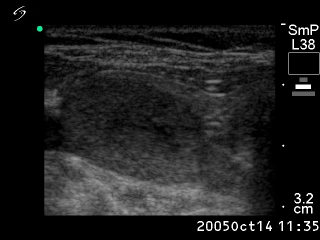
Ultrasonography: the thyroids were echonormal. There was a solitary nodule in the right lobe. A halo sign was not seen, but perinodular blood flow was present. The dimensions of the nodule were 25x16x27 mm.
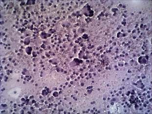
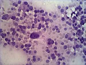
Cytological picture: there is no colloid on the smear. A very cellular picture - epithelial cells with abundant cytoplasm and eccentric nuclei with prominent nucleoli are on the smear. Most of the cells are dissociated. Note the many triangular forms, the great number of atypical enlarged and multinucleated cells. On this pattern the possibility of a Hürthle-cell tumor and a medullary cancer must be considered. The presence of several small cell clusters corresponding to microfollicles and that of colloid favor the former possibility.
Cytological diagnosis: Hürthle cell tumor.
Histopathological diagnosis: Hürthle cell adenoma
Comment: the clues in differentiation from medullary cancer are the presence of microfollicles on the smear and the perinodular blood flow seen on ultrasonography.