
|
|
Oxyphilic adenoma - Case 17.
|
|


Clinical presentation: a 53-year-old woman was referred for an evaluation of a thyroid nodule that had been discovered on carotid Doppler examination.
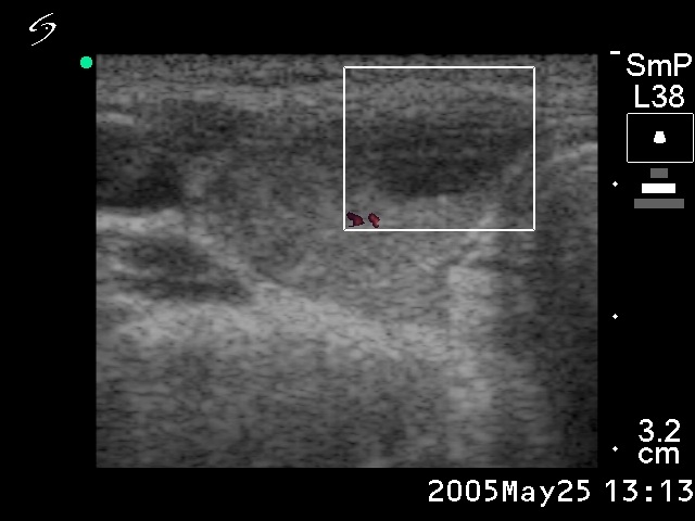
Palpation: a solitary nodule in the right lobe.
Hormonal examination: indicated euthyroidism with TSH-level 1.94 mIU/L, and FT4 15.2 pM/L.
Ultrasonography: the thyroid was minimally hypoechogenic. Two hypoechogenic lesions were found in the right lobe. The larger one contained microcalcifications. There was neither perinodular nor intranodular blood flow.
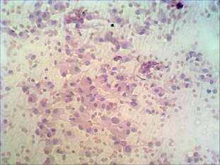
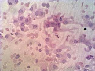
Cytology: there was no colloid in the background. Thyrocytes were found dissociated and in several trabecular structures. Follicular cells exhibited oxyphilic metaplasia. Great proportion of them contained inclusion. Scattered number of lymphocytes were also found.
FNAC report: suspicion of oxyphilic variant of papillary cancer.
Histopathological diagnosis: benign oxyphilic adenoma and chronic lymphocytic thyroiditis outside the nodule.
Comments:
-
The presence of inclusion in oxyphilic cells compared with that in non-metaplastic follicular cells has much less specificity for papillary cancer.
-
This case represents the limitations of FNAC. Such cytological pattern requires surgery because it is identical with that observed in oxyphilic variant of papillary cancer.