
|
|
Differentiation of cystic thyroid lesions - Case 142doi: 10.24390/thyrocase142.00
|

|


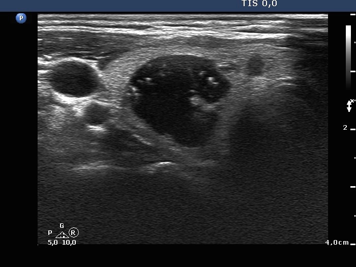
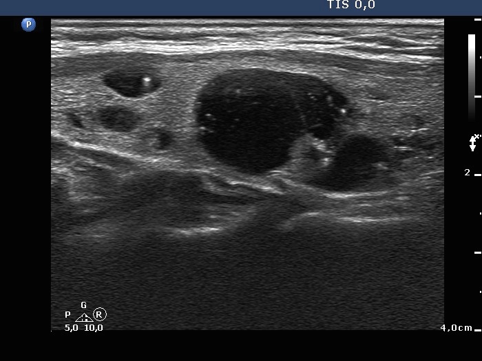
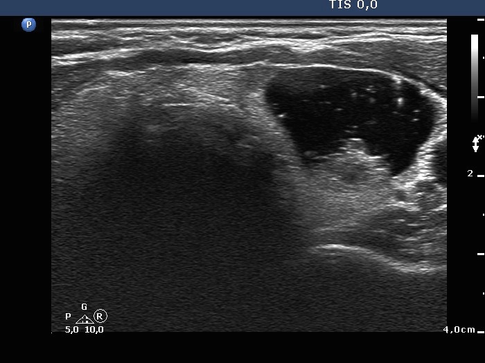
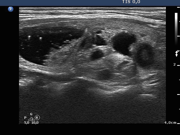

First examination (first row of images)
Clinical presentation. A 44-year-old woman has noticed the enlargement of the region of the thyroid for two months.
Palpation. Both lobes were nodular on palpation.
Result of blood tests: TSH 1.28 mIU/L.
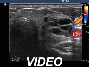
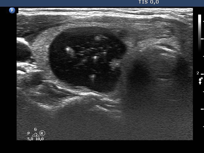
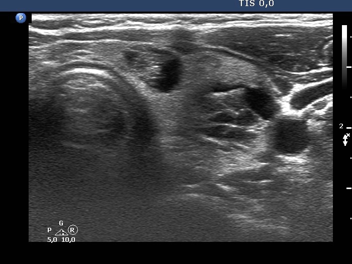
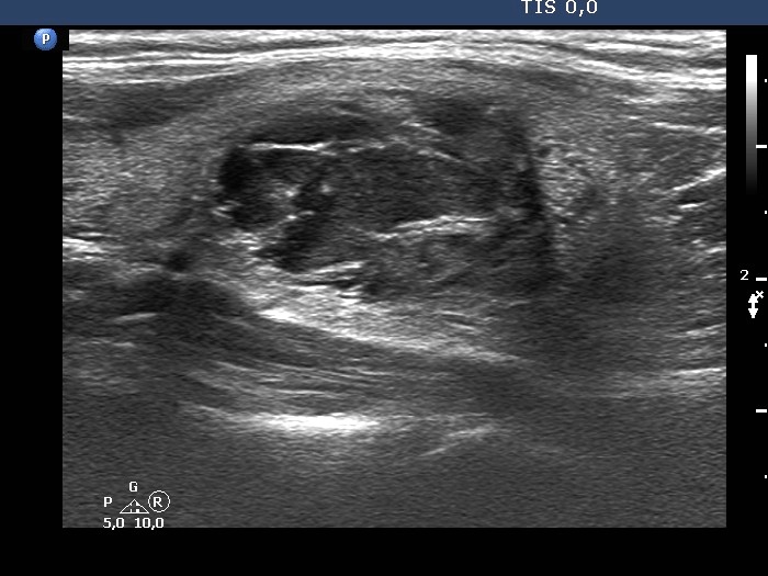
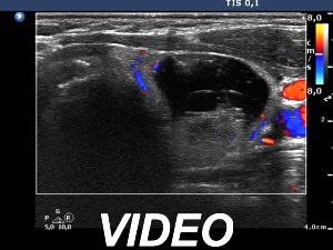
Ultrasonography. The thyroid was echonormal. There were multiple cystic areas and nodules in both lobes. The smaller cystic lesions in the right lobe were partly cystic areas, partly doubtful cystic lesions. The largest lesion was a cystic nodule with a difficult-to-categorize pattern. The nodule in the upper part of the left lobe was a peripheral-type cyst, another lesion under the previous one was a spongiform-type nodule.We aspirated 2.8 mL brown fluid from the peripheral type cyst, thereafter made an US-guided aspiration from the solid part. Aspiration cytology resulted in benign cystic lesion.
Suggestion: thyroid ultrasound in a year.
Second examination 3 years later (second row of images)
Clinical presentation. The patient had no compaints.
Palpation: unchanged.
Result of blood tests: TSH 2.44 mIU/L.
Ultrasonography. The presentation remained unchanged except for two conditions. Firstly, the cystic nodule in the right lobe has slightly increased while the cystic nodule in the upper part of the left lobe has significantly decreased, i.e. it has not refilled.Suggestion: ultrasound in three years.


