
|
|
Intranodular hyperechogenic figures - case 1429doi: 10.24390/thyrocase1429.00
|
|


Clinical presentation: A 52-year-old woman was referred for aspiration cytology. A nodular goiter was discovered in the right thyroid on PET CT scan performed on follow-up of malignant melanoma.
Palpation: no abnormality.
Functional state: euthyroidism with TSH 1.15 mIU/L.
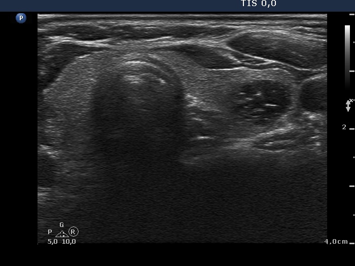
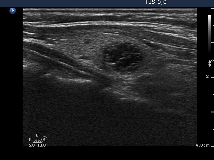
Ultrasonography. The thyroid was echonormal. There was a minimally-moderately hypoechogenic nodule in the right while a cystic nodule in the left lobe. The latter presented several intranodular hyperechogenic figures.
Both lesions were aspirated and cytology resulted in both cases in benign lesion.
Comment. Neither intranodular figures are typical comet-tail artifacts. Nevertheless, the size of the figures, the cystic pattern of the nodule stands for comet-tail artifacts.