
The role of complex diagnosis - other examples - Case 2. (ultrasonographic picture 4)
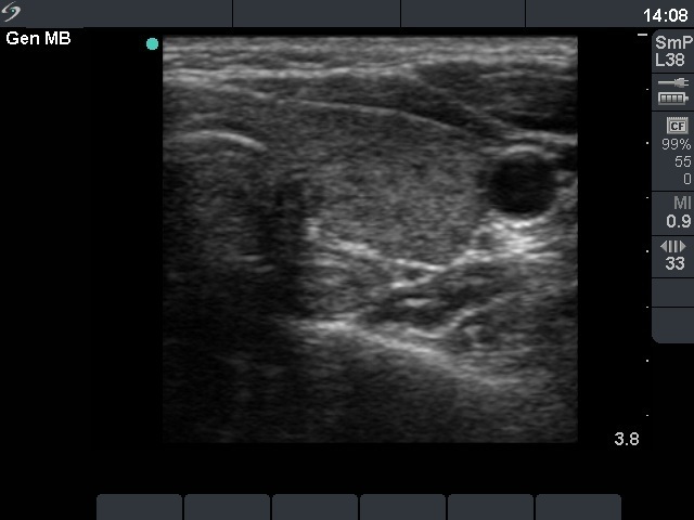 Left lobe, horizontal scan. This lobe has a similar presentation: there are minimally hypoechogenic areas within an echonormal background. |
The role of complex diagnosis - other examples - Case 2. (ultrasonographic picture 4)
Left lobe, horizontal scan. This lobe has a similar presentation: there are minimally hypoechogenic areas within an echonormal background. |
